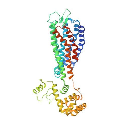
5TZY

GPR40 in complex with AgoPAM AP8 and partial agonist MK-8666


Experimental Data Snapshot

  • Method: X-RAY DIFFRACTION
  • Resolution: 3.22 Å
  • R-Value Free: 0.287 
  • R-Value Work: 0.269 
  • R-Value Observed: 0.270 

Starting Model: experimental
View more details

wwPDB Validation   3D Report Full Report


Ligand Structure Quality Assessment 


This is version 1.3 of the entry. See complete history


Literature

Structural basis for the cooperative allosteric activation of the free fatty acid receptor GPR40.

Lu, J.Byrne, N.Wang, J.Bricogne, G.Brown, F.K.Chobanian, H.R.Colletti, S.L.Di Salvo, J.Thomas-Fowlkes, B.Guo, Y.Hall, D.L.Hadix, J.Hastings, N.B.Hermes, J.D.Ho, T.Howard, A.D.Josien, H.Kornienko, M.Lumb, K.J.Miller, M.W.Patel, S.B.Pio, B.Plummer, C.W.Sherborne, B.S.Sheth, P.Souza, S.Tummala, S.Vonrhein, C.Webb, M.Allen, S.J.Johnston, J.M.Weinglass, A.B.Sharma, S.Soisson, S.M.

(2017) Nat Struct Mol Biol 24: 570-577

  • DOI: https://doi.org/10.1038/nsmb.3417
  • Primary Citation of Related Structures:  
    5TZR, 5TZY

  • PubMed Abstract: 

    Clinical studies indicate that partial agonists of the G-protein-coupled, free fatty acid receptor 1 GPR40 enhance glucose-dependent insulin secretion and represent a potential mechanism for the treatment of type 2 diabetes mellitus. Full allosteric agonists (AgoPAMs) of GPR40 bind to a site distinct from partial agonists and can provide additional efficacy. We report the 3.2-Å crystal structure of human GPR40 (hGPR40) in complex with both the partial agonist MK-8666 and an AgoPAM, which exposes a novel lipid-facing AgoPAM-binding pocket outside the transmembrane helical bundle. Comparison with an additional 2.2-Å structure of the hGPR40-MK-8666 binary complex reveals an induced-fit conformational coupling between the partial agonist and AgoPAM binding sites, involving rearrangements of the transmembrane helices 4 and 5 (TM4 and TM5) and transition of the intracellular loop 2 (ICL2) into a short helix. These conformational changes likely prime GPR40 to a more active-like state and explain the binding cooperativity between these ligands.


  • Organizational Affiliation

    Department of Structural Chemistry, Merck Research Laboratories, West Point, Pennsylvania, USA.


Macromolecules
Find similar proteins by:  (by identity cutoff)  |  3D Structure
Entity ID: 1
MoleculeChains Sequence LengthOrganismDetailsImage
Free fatty acid receptor 1,Endolysin,Free fatty acid receptor 1491Homo sapiensTequatrovirus T4Mutation(s): 7 
Gene Names: FFAR1GPR40
EC: 3.2.1.17
Membrane Entity: Yes 
UniProt & NIH Common Fund Data Resources
Find proteins for P00720 (Enterobacteria phage T4)
Explore P00720 
Go to UniProtKB:  P00720
Find proteins for O14842 (Homo sapiens)
Explore O14842 
Go to UniProtKB:  O14842
GTEx:  ENSG00000126266 
Entity Groups  
Sequence Clusters30% Identity50% Identity70% Identity90% Identity95% Identity100% Identity
UniProt GroupsP00720O14842
Sequence Annotations
Expand
  • Reference Sequence
Small Molecules
Ligands 3 Unique
IDChains Name / Formula / InChI Key2D Diagram3D Interactions
7OS
Query on 7OS

Download Ideal Coordinates CCD File 
D [auth A](2S,3R)-3-cyclopropyl-3-[(2R)-2-(1-{(1S)-1-[5-fluoro-2-(trifluoromethoxy)phenyl]ethyl}piperidin-4-yl)-3,4-dihydro-2H-1-benzopyran-7-yl]-2-methylpropanoic acid
C30 H35 F4 N O4
ADYYYLTWZYYGNX-LJYIQKJHSA-N
MK6
Query on MK6

Download Ideal Coordinates CCD File 
C [auth A](5aR,6S,6aS)-3-({2',6'-dimethyl-4'-[3-(methylsulfonyl)propoxy][1,1'-biphenyl]-3-yl}methoxy)-5,5a,6,6a-tetrahydrocyclopropa[4,5]cyclopenta[1,2-c]pyridine-6-carboxylic acid
C29 H31 N O6 S
CODQKEMYZZKQAE-QPVYNBJUSA-N
OLC
Query on OLC

Download Ideal Coordinates CCD File 
B [auth A](2R)-2,3-dihydroxypropyl (9Z)-octadec-9-enoate
C21 H40 O4
RZRNAYUHWVFMIP-GDCKJWNLSA-N
Binding Affinity Annotations 
IDSourceBinding Affinity
MK6 BindingDB:  5TZY EC50: 5.6 (nM) from 1 assay(s)
Experimental Data & Validation

Experimental Data

  • Method: X-RAY DIFFRACTION
  • Resolution: 3.22 Å
  • R-Value Free: 0.287 
  • R-Value Work: 0.269 
  • R-Value Observed: 0.270 
  • Space Group: P 1 21 1
Unit Cell:
Length ( Å )Angle ( ˚ )
a = 53.88α = 90
b = 63.95β = 90.52
c = 90.96γ = 90
Software Package:
Software NamePurpose
BUSTERrefinement
XDSdata reduction
autoPROCdata scaling
PHASERphasing

Structure Validation

View Full Validation Report


Ligand Structure Quality Assessment 


Entry History 

Deposition Data

Revision History  (Full details and data files)

  • Version 1.0: 2017-06-07
    Type: Initial release
  • Version 1.1: 2017-06-21
    Changes: Database references
  • Version 1.2: 2017-07-19
    Changes: Database references
  • Version 1.3: 2023-10-04
    Changes: Data collection, Database references, Refinement description