Asp263 missense variants perturb the active site of human phosphoglucomutase 1.
Stiers, K.M., Graham, A.C., Kain, B.N., Beamer, L.J.(2017) FEBS J 284: 937-947
- PubMed: 28117557 Search on PubMedSearch on PubMed Central
- DOI: https://doi.org/10.1111/febs.14025
- Primary Citation Related Structures:
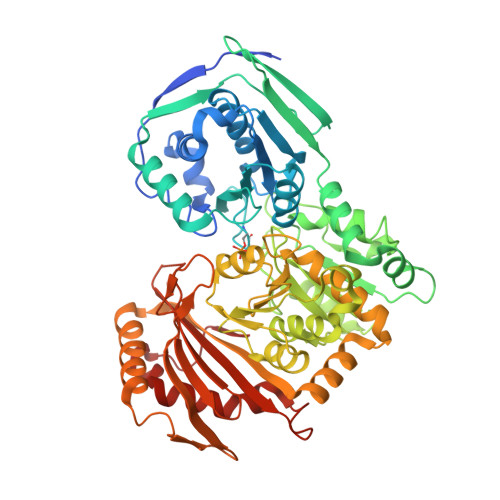
5JN5, 5TR2 - PubMed Abstract:
The enzyme phosphoglucomutase 1 (PGM1) plays a central role in glucose homeostasis. Clinical studies have identified mutations in human PGM1 as the cause of PGM1 deficiency, an inherited metabolic disease. One residue, Asp263, has two known variants associated with disease: D263G and D263Y. Biochemical studies have shown that these mutants are soluble and well folded, but have significant catalytic impairment. To better understand this catalytic defect, we determined crystal structures of these two missense variants, both of which reveal a similar and indirect structural change due to the loss of a conserved salt bridge between Asp263 and Arg293. The arginine reorients into the active site, making interactions with residues responsible for substrate binding. Biochemical studies also show that the catalytic phosphoserine of the missense variants is more stable to hydrolysis relative to wild-type enzyme. The structural perturbation resulting from mutation of this single amino acid reveals the molecular mechanism underlying PGM1 deficiency in these missense variants. Structural data are available in the PDB under the accession numbers 5JN5 and 5TR2.
- Biochemistry Department, University of Missouri, Columbia, MO, USA.
Organizational Affiliation: