3D Printed Micrometer-Scale Polymer Mounts for Single Crystal Analysis.
Macdonald, N.P., Bunton, G.L., Park, A.Y., Breadmore, M.C., Kilah, N.L.(2017) Anal Chem 89: 4405-4408
- PubMed: 28319372 Search on PubMed
- DOI: https://doi.org/10.1021/acs.analchem.7b00443
- Primary Citation Related Structures:
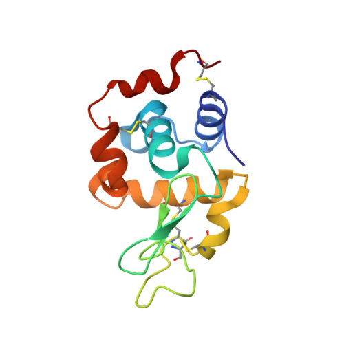
5TK0 - PubMed Abstract:
3D printed micrometer-scale polymer mounts for single crystal analysis have been prepared by photopolymerization using digital light projection stereolithography (DLP-SLA), with a commercially available digital light projection stereolithography printer (US$4000) and 3DM-ABS resin (US$150 per liter). The polymer mounts were prepared in batches of 49 in 1 h 15 min, which allowed for rapid prototyping and testing of new crystal mounting designs, with a resin cost of 0.2¢ US per mount. The suitability of the 3D printed mounts for single crystal crystallography has been demonstrated through their use in Cu Kα X-ray diffraction experiments of Rochelle salt (sodium potassium tartrate), the protein lysozyme, and has been employed for routine crystallographic analysis of organic and inorganic materials.
- School of Physical Sciences - Chemistry, ‡ARC Centre of Excellence for Electromaterials Science, and §Australian Centre for Research on Separation Science (ACROSS), University of Tasmania , Dobson Road, Sandy Bay, Tasmania 7005, Australia.
Organizational Affiliation: