5TGL

A MODEL FOR INTERFACIAL ACTIVATION IN LIPASES FROM THE STRUCTURE OF A FUNGAL LIPASE-INHIBITOR COMPLEX


Experimental Data Snapshot

  • Method: X-RAY DIFFRACTION
  • Resolution: 3.00 Å
  • R-Value Observed: 0.185 

wwPDB Validation   3D Report Full Report


This is version 1.3 of the entry. See complete history


Literature

A model for interfacial activation in lipases from the structure of a fungal lipase-inhibitor complex.

Brzozowski, A.M.Derewenda, U.Derewenda, Z.S.Dodson, G.G.Lawson, D.M.Turkenburg, J.P.Bjorkling, F.Huge-Jensen, B.Patkar, S.A.Thim, L.

(1991) Nature 351: 491-494

  • DOI: https://doi.org/10.1038/351491a0
  • Primary Citation of Related Structures:  
    5TGL

  • PubMed Abstract: 

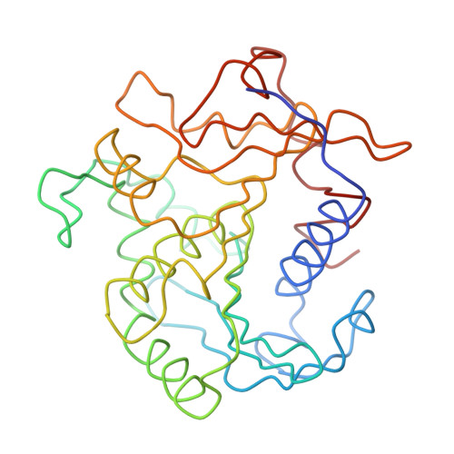
    Lipases are hydrolytic enzymes which break down triacylglycerides into free fatty acids and glycerols. They have been classified as serine hydrolases owing to their inhibition by diethyl p-nitrophenyl phosphate. Lipase activity is greatly increased at the lipid-water interface, a phenomenon known as interfacial activation. X-ray analysis has revealed the atomic structures of two triacylglycerol lipases, unrelated in sequence: the human pancreatic lipase (hPL)4, and an enzyme isolated from the fungus Rhizomucor (formerly Mucor) miehei (RmL). In both enzymes the active centres contain structurally analogous Asp-His-Ser triads (characteristic of serine proteinases), which are buried completely beneath a short helical segment, or 'lid'. Here we present the crystal structure (at 3 A resolution) of a complex of R. miehei lipase with n-hexylphosphonate ethyl ester in which the enzyme's active site is exposed by the movement of the helical lid. This movement also increases the nonpolarity of the surface surrounding the catalytic site. We propose that the structure of the enzyme in this complex is equivalent to the activated state generated by the oil-water interface.


  • Organizational Affiliation

    Department of Chemistry, University of York, Heslington, UK.


Macromolecules
Find similar proteins by:  (by identity cutoff)  |  3D Structure
Entity ID: 1
MoleculeChains Sequence LengthOrganismDetailsImage
LIPASE269Rhizomucor mieheiMutation(s): 0 
EC: 3.1.1.3
UniProt
Find proteins for P19515 (Rhizomucor miehei)
Explore P19515 
Go to UniProtKB:  P19515
Entity Groups  
Sequence Clusters30% Identity50% Identity70% Identity90% Identity95% Identity100% Identity
UniProt GroupP19515
Sequence Annotations
Expand
  • Reference Sequence
Small Molecules
Ligands 1 Unique
IDChains Name / Formula / InChI Key2D Diagram3D Interactions
HEE
Query on HEE

Download Ideal Coordinates CCD File 
B [auth A]N-HEXYLPHOSPHONATE ETHYL ESTER
C8 H19 O3 P
XPLOQMVUXWZLET-UHFFFAOYSA-N
Experimental Data & Validation

Experimental Data

  • Method: X-RAY DIFFRACTION
  • Resolution: 3.00 Å
  • R-Value Observed: 0.185 
  • Space Group: C 2 2 21
Unit Cell:
Length ( Å )Angle ( ˚ )
a = 48.3α = 90
b = 93.9β = 90
c = 122.1γ = 90
Software Package:
Software NamePurpose
X-PLORmodel building
PROLSQrefinement
X-PLORrefinement
X-PLORphasing

Structure Validation

View Full Validation Report


Entry History 

Revision History  (Full details and data files)

  • Version 1.0: 1994-01-31
    Type: Initial release
  • Version 1.1: 2008-03-03
    Changes: Version format compliance
  • Version 1.2: 2011-07-13
    Changes: Version format compliance
  • Version 1.3: 2024-03-06
    Changes: Data collection, Database references, Derived calculations, Other