5TF6 | pdb_00005tf6

Structure and conformational plasticity of the U6 small nuclear ribonucleoprotein core


Experimental Data Snapshot

  • Method: X-RAY DIFFRACTION
  • Resolution: 2.30 Å
  • R-Value Free: 
    0.232 (Depositor), 0.234 (DCC) 
  • R-Value Work: 
    0.185 (Depositor), 0.190 (DCC) 
  • R-Value Observed: 
    0.186 (Depositor) 

wwPDB Validation 3D Report Full Report

Validation slider image for 5TF6

This is version 1.4 of the entry. See complete history

Literature

Structure and conformational plasticity of the U6 small nuclear ribonucleoprotein core.

Montemayor, E.J.Didychuk, A.L.Liao, H.Hu, P.Brow, D.A.Butcher, S.E.

(2017) Acta Crystallogr D Struct Biol 73: 1-8

  • DOI: https://doi.org/10.1107/S2059798316018222
  • Primary Citation Related Structures: 
    5TF6

  • PubMed Abstract: 

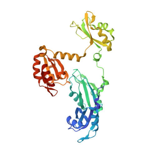
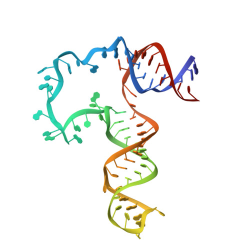
    U6 small nuclear RNA (snRNA) is a key component of the active site of the spliceosome, a large ribonucleoprotein complex that catalyzes the splicing of precursor messenger RNA. Prior to its incorporation into the spliceosome, U6 is bound by the protein Prp24, which facilitates unwinding of the U6 internal stem-loop (ISL) so that it can pair with U4 snRNA. A previously reported crystal structure of the `core' of the U6 small nuclear ribonucleoprotein (snRNP) contained an ISL-stabilized A62G mutant of U6 bound to all four RNA-recognition motif (RRM) domains of Prp24 [Montemayor et al. (2014), Nature Struct. Mol. Biol. 21, 544-551]. The structure revealed a novel topology containing interlocked rings of protein and RNA that was not predicted by prior biochemical and genetic data. Here, the crystal structure of the U6 snRNP core with a wild-type ISL is reported. This complex crystallized in a new space group, apparently owing in part to the presence of an intramolecular cross-link in RRM1 that was not observed in the previously reported U6-A62G structure. The structure exhibits the same protein-RNA interface and maintains the unique interlocked topology. However, the orientation of the wild-type ISL is altered relative to the A62G mutant structure, suggesting inherent structural dynamics that may facilitate its pairing with U4. Consistent with their similar architectures in the crystalline state, the wild-type and A62G variants of U6 exhibit similar Prp24-binding affinities and electrophoretic mobilities when analyzed by gel-shift assay.


  • Organizational Affiliation
    • Department of Biochemistry, University of Wisconsin-Madison, Madison, WI 53706, USA.

Macromolecule Content 

  • Total Structure Weight: 133.63 kDa 
  • Atom Count: 9,375 
  • Modeled Residue Count: 856 
  • Deposited Residue Count: 892 
  • Unique protein chains: 1
  • Unique nucleic acid chains: 1

Macromolecules


Find similar proteins by:|  3D Structure
Entity ID: 1
MoleculeChains  Sequence LengthOrganismDetailsImage
U4/U6 snRNA-associated-splicing factor PRP24
A, C
374Saccharomyces cerevisiae S288CMutation(s): 0 
Gene Names: PRP24YMR268CYM8156.10C
UniProt
Find proteins for P49960 (Saccharomyces cerevisiae (strain ATCC 204508 / S288c))
Explore P49960 
Go to UniProtKB:  P49960
Entity Groups
Sequence Clusters30% Identity50% Identity70% Identity90% Identity95% Identity100% Identity
UniProt GroupP49960
Sequence Annotations
Expand
Reference Sequence
Find similar nucleic acids by:  (by identity cutoff) 
Entity ID: 2
MoleculeChains LengthOrganismImage
U6 snRNA
B, D
72Saccharomyces cerevisiae
Sequence Annotations
Expand
Reference Sequence

Small Molecules

Ligands 3 Unique
IDChains Name / Formula / InChI Key2D Diagram3D Interactions
GOL

Query on GOL


Download:Ideal Coordinates CCD File
H [auth A],
I [auth A],
K [auth B],
P [auth C],
Q [auth C]
GLYCEROL
C3 H8 O3
PEDCQBHIVMGVHV-UHFFFAOYSA-N
K

Query on K


Download:Ideal Coordinates CCD File
E [auth A],
J [auth B],
L [auth C],
M [auth C],
R [auth D]
POTASSIUM ION
K
NPYPAHLBTDXSSS-UHFFFAOYSA-N
CL

Query on CL


Download:Ideal Coordinates CCD File
F [auth A],
G [auth A],
N [auth C],
O [auth C]
CHLORIDE ION
Cl
VEXZGXHMUGYJMC-UHFFFAOYSA-M

Experimental Data & Validation

Experimental Data

  • Method: X-RAY DIFFRACTION
  • Resolution: 2.30 Å
  • R-Value Free:  0.232 (Depositor), 0.234 (DCC) 
  • R-Value Work:  0.185 (Depositor), 0.190 (DCC) 
  • R-Value Observed: 0.186 (Depositor) 
Space Group: P 21 21 21
Unit Cell:
Length ( Å )Angle ( ˚ )
a = 76.921α = 90
b = 84.446β = 90
c = 255.142γ = 90
Software Package:
Software NamePurpose
Aimlessdata scaling
PHENIXrefinement
PDB_EXTRACTdata extraction
XDSdata reduction
PHASERphasing

Structure Validation

View Full Validation Report


Entry History 

& Funding Information

Deposition Data


Funding OrganizationLocationGrant Number
National Institutes of Health/National Institute of General Medical Sciences (NIH/NIGMS)United StatesR01 GM065166
National Institutes of Health/National Institute of General Medical Sciences (NIH/NIGMS)United StatesR35 GM118131
National Institutes of Health/National Institute of General Medical Sciences (NIH/NIGMS)United StatesR35 GM118075

Revision History  (Full details and data files)

  • Version 1.0: 2017-01-11
    Type: Initial release
  • Version 1.1: 2017-01-18
    Changes: Database references
  • Version 1.2: 2017-09-27
    Changes: Author supporting evidence
  • Version 1.3: 2019-12-25
    Changes: Author supporting evidence
  • Version 1.4: 2024-03-06
    Changes: Data collection, Database references, Derived calculations