5T3B

Crystal structure of BpGH50


Experimental Data Snapshot

  • Method: X-RAY DIFFRACTION
  • Resolution: 1.40 Å
  • R-Value Free: 0.145 
  • R-Value Work: 0.106 
  • R-Value Observed: 0.108 

wwPDB Validation   3D Report Full Report


This is version 1.5 of the entry. See complete history


Literature

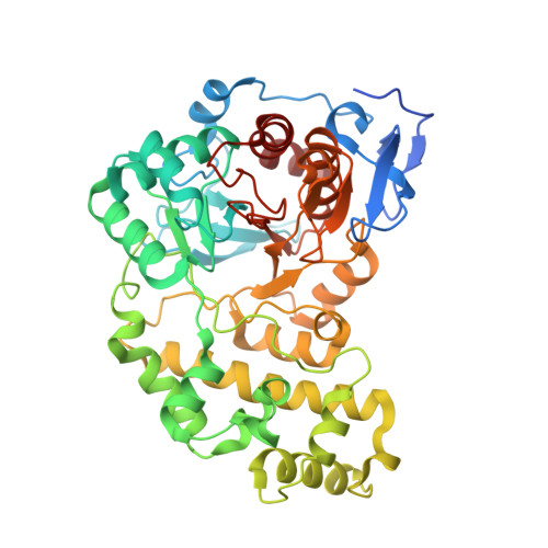
Structure of a glycoside hydrolase family 50 enzyme from a subfamily that is enriched in human gut microbiome bacteroidetes.

Giles, K.Pluvinage, B.Boraston, A.B.

(2017) Proteins 85: 182-187

  • DOI: https://doi.org/10.1002/prot.25189
  • Primary Citation of Related Structures:  
    5T3B

  • PubMed Abstract: 

    The polysaccharide utilization locus in Bacteroides plebeius that confers the ability to catabolize porphyran contains a putative GH50 β-agarase (BACPLE_01683, BpGH50). BpGH50 did not show any clear activity on agarose or on the related algal galactans porphyran and carrageenan. However, the 1.4 Å resolution X-ray crystal structure of BpGH50 confirmed its possession of the core (α/β) 8 barrel fold found in GH50 enzymes as well as the structural conservation of the catalytic residues and some substrate binding residues. Examination of the structure supports assignment of this protein as a β-galactosidase but suggests that it may utilize a different, possibly hybrid, algal galactan substrate. Proteins 2016; 85:182-187. © 2016 Wiley Periodicals, Inc.


  • Organizational Affiliation

    Biochemistry & Microbiology, University of Victoria, PO Box 3055 STN CSC, Victoria, British Columbia V8W 3P6, Canada.


Macromolecules
Find similar proteins by:  (by identity cutoff)  |  3D Structure
Entity ID: 1
MoleculeChains Sequence LengthOrganismDetailsImage
Glycoside Hydrolase
A, B
481Phocaeicola plebeiusMutation(s): 0 
Gene Names: BACPLE_01683
UniProt
Find proteins for B5CY86 (Phocaeicola plebeius (strain DSM 17135 / JCM 12973 / M2))
Explore B5CY86 
Go to UniProtKB:  B5CY86
Entity Groups  
Sequence Clusters30% Identity50% Identity70% Identity90% Identity95% Identity100% Identity
UniProt GroupB5CY86
Sequence Annotations
Expand
  • Reference Sequence
Small Molecules
Ligands 1 Unique
IDChains Name / Formula / InChI Key2D Diagram3D Interactions
EDO
Query on EDO

Download Ideal Coordinates CCD File 
C [auth A]
D [auth A]
E [auth A]
F [auth A]
G [auth A]
C [auth A],
D [auth A],
E [auth A],
F [auth A],
G [auth A],
H [auth A],
I [auth A],
J [auth A],
K [auth A],
L [auth A],
M [auth A],
N [auth A],
O [auth A],
P [auth A],
Q [auth B],
R [auth B],
S [auth B],
T [auth B]
1,2-ETHANEDIOL
C2 H6 O2
LYCAIKOWRPUZTN-UHFFFAOYSA-N
Experimental Data & Validation

Experimental Data

  • Method: X-RAY DIFFRACTION
  • Resolution: 1.40 Å
  • R-Value Free: 0.145 
  • R-Value Work: 0.106 
  • R-Value Observed: 0.108 
  • Space Group: P 1 21 1
Unit Cell:
Length ( Å )Angle ( ˚ )
a = 73.1α = 90
b = 87.37β = 105.5
c = 74.67γ = 90
Software Package:
Software NamePurpose
MOSFLMdata reduction
SCALAdata scaling
PHASERphasing
REFMACrefinement
PDB_EXTRACTdata extraction

Structure Validation

View Full Validation Report


Entry History & Funding Information

Deposition Data


Funding OrganizationLocationGrant Number
Natural Sciences and Engineering Research Council (NSERC, Canada)Canada--

Revision History  (Full details and data files)

  • Version 1.0: 2016-10-12
    Type: Initial release
  • Version 1.1: 2016-11-02
    Changes: Database references
  • Version 1.2: 2017-01-04
    Changes: Database references
  • Version 1.3: 2017-09-27
    Changes: Author supporting evidence
  • Version 1.4: 2020-01-08
    Changes: Author supporting evidence
  • Version 1.5: 2024-03-06
    Changes: Data collection, Database references