5SX2

Crystal structure of the D141E mutant of B. pseudomallei KatG at pH 8.0.


Experimental Data Snapshot

  • Method: X-RAY DIFFRACTION
  • Resolution: 2.15 Å
  • R-Value Free: 0.180 
  • R-Value Work: 0.142 
  • R-Value Observed: 0.144 

wwPDB Validation   3D Report Full Report


Ligand Structure Quality Assessment 


This is version 1.4 of the entry. See complete history


Literature

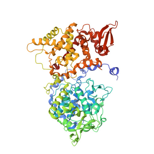
Two alternative substrate paths for compound I formation and reduction in catalase-peroxidase KatG from Burkholderia pseudomallei.

Deemagarn, T.Wiseman, B.Carpena, X.Ivancich, A.Fita, I.Loewen, P.C.

(2007) Proteins 66: 219-228

  • DOI: https://doi.org/10.1002/prot.21209
  • Primary Citation of Related Structures:  
    5SX1, 5SX2

  • PubMed Abstract: 

    Five residues in the multifunctional catalase-peroxidase KatG of Burkholderia pesudomallei are essential for catalase, but not peroxidase, activity. Asp141 is the only one of these catalase-specific residues not related with the covalent adduct found in KatGs that when replaced with a nonacidic residue reduces catalase activity to 5% of native levels. Replacing the nearby catalytic residue Arg108 causes a reduction in catalase activity to 35% of native levels, whereas a variant with both Asp141 and Arg108 replaced exhibits near normal catalase activity (82% of native), suggesting a synergism in the roles of the two residues in support of catalase activity in the enzyme. Among the Asp141 variants, D141E is unique in retaining normal catalase activity but with modified kinetics, suggesting more favorable compound I formation and less favorable compound I reduction. The crystal structure of the D141E variant has been determined at 1.8-A resolution, revealing that the carboxylate of Glu141 is moved only slightly compared with Asp141, but retains its hydrogen bond interaction with the main chain nitrogen of Ile237. In contrast, the low temperature ferric Electron Paramagnetic Resonance spectra of the D141A, R108A, and R108A/D141A variants are consistent with modifications of the water matrix and/or the relative positioning of the distal residue side chains. Such changes explain the reduction in catalase activity in all but the double variant R108A/D141A. Two pathways of hydrogen bonded solvent lead from the entrance channel into the heme active site, one running between Asp141 and Arg108 and the second between Asp141 and the main chain atoms of residues 237-239. It is proposed that binding of substrate H(2)O(2) to Asp141 and Arg108 controls H(2)O(2) access to the heme active site, thereby modulating the catalase reaction.


  • Organizational Affiliation

    Department of Microbiology, University of Manitoba, Winnipeg, Manitoba R3T 2N2, Canada.


Macromolecules
Find similar proteins by:  (by identity cutoff)  |  3D Structure
Entity ID: 1
MoleculeChains Sequence LengthOrganismDetailsImage
Catalase-peroxidase
A, B
728Burkholderia pseudomallei 1710bMutation(s): 1 
Gene Names: katGBURPS1710b_3366
EC: 1.11.1.21
UniProt
Find proteins for Q3JNW6 (Burkholderia pseudomallei (strain 1710b))
Explore Q3JNW6 
Go to UniProtKB:  Q3JNW6
Entity Groups  
Sequence Clusters30% Identity50% Identity70% Identity90% Identity95% Identity100% Identity
UniProt GroupQ3JNW6
Sequence Annotations
Expand
  • Reference Sequence
Small Molecules
Ligands 6 Unique
IDChains Name / Formula / InChI Key2D Diagram3D Interactions
HEM
Query on HEM

Download Ideal Coordinates CCD File 
C [auth A],
K [auth B]
PROTOPORPHYRIN IX CONTAINING FE
C34 H32 Fe N4 O4
KABFMIBPWCXCRK-RGGAHWMASA-L
TRS
Query on TRS

Download Ideal Coordinates CCD File 
I [auth A],
P [auth B]
2-AMINO-2-HYDROXYMETHYL-PROPANE-1,3-DIOL
C4 H12 N O3
LENZDBCJOHFCAS-UHFFFAOYSA-O
MPD
Query on MPD

Download Ideal Coordinates CCD File 
G [auth A],
H [auth A],
O [auth B]
(4S)-2-METHYL-2,4-PENTANEDIOL
C6 H14 O2
SVTBMSDMJJWYQN-YFKPBYRVSA-N
PO4
Query on PO4

Download Ideal Coordinates CCD File 
J [auth A],
Q [auth B]
PHOSPHATE ION
O4 P
NBIIXXVUZAFLBC-UHFFFAOYSA-K
OXY
Query on OXY

Download Ideal Coordinates CCD File 
E [auth A],
F [auth A],
M [auth B],
N [auth B]
OXYGEN MOLECULE
O2
MYMOFIZGZYHOMD-UHFFFAOYSA-N
NA
Query on NA

Download Ideal Coordinates CCD File 
D [auth A],
L [auth B]
SODIUM ION
Na
FKNQFGJONOIPTF-UHFFFAOYSA-N
Modified Residues  1 Unique
IDChains TypeFormula2D DiagramParent
TOX
Query on TOX
A, B
L-PEPTIDE LINKINGC11 H12 N2 O4TRP
Experimental Data & Validation

Experimental Data

  • Method: X-RAY DIFFRACTION
  • Resolution: 2.15 Å
  • R-Value Free: 0.180 
  • R-Value Work: 0.142 
  • R-Value Observed: 0.144 
  • Space Group: P 21 21 21
Unit Cell:
Length ( Å )Angle ( ˚ )
a = 100.327α = 90
b = 114.824β = 90
c = 174.31γ = 90
Software Package:
Software NamePurpose
REFMACrefinement
PDB_EXTRACTdata extraction
DENZOdata reduction
SCALEPACKdata scaling
MOLREPphasing

Structure Validation

View Full Validation Report


Ligand Structure Quality Assessment 


Entry History & Funding Information

Deposition Data

  • Released Date: 2016-09-07 
  • Deposition Author(s): Loewen, P.C.
  • This entry supersedes: 2DV2

Funding OrganizationLocationGrant Number
Natural Sciences and Engineering Research Council (NSERC, Canada)CanadaD9600

Revision History  (Full details and data files)

  • Version 1.0: 2016-09-07
    Type: Initial release
  • Version 1.1: 2017-09-27
    Changes: Author supporting evidence, Derived calculations, Structure summary
  • Version 1.2: 2020-01-08
    Changes: Author supporting evidence
  • Version 1.3: 2023-10-04
    Changes: Data collection, Database references, Derived calculations, Refinement description
  • Version 1.4: 2023-11-15
    Changes: Data collection