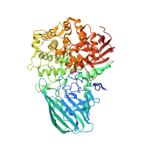
5SWI

Crystal structure of SpGH92 in complex with mannose


Experimental Data Snapshot

  • Method: X-RAY DIFFRACTION
  • Resolution: 2.15 Å
  • R-Value Free: 0.221 
  • R-Value Work: 0.197 
  • R-Value Observed: 0.198 

wwPDB Validation   3D Report Full Report


Ligand Structure Quality Assessment 


This is version 1.4 of the entry. See complete history


Literature

Molecular Characterization of N-glycan Degradation and Transport in Streptococcus pneumoniae and Its Contribution to Virulence.

Robb, M.Hobbs, J.K.Woodiga, S.A.Shapiro-Ward, S.Suits, M.D.McGregor, N.Brumer, H.Yesilkaya, H.King, S.J.Boraston, A.B.

(2017) PLoS Pathog 13: e1006090-e1006090

  • DOI: https://doi.org/10.1371/journal.ppat.1006090
  • Primary Citation of Related Structures:  
    5SUO, 5SWA, 5SWB, 5SWI

  • PubMed Abstract: 
  • The carbohydrate-rich coating of human tissues and cells provide a first point of contact for colonizing and invading bacteria. Commensurate with N-glycosylation being an abundant form of protein glycosylation that has critical functional roles in the host, some host-adapted bacteria possess the machinery to process N-linked glycans ...

    The carbohydrate-rich coating of human tissues and cells provide a first point of contact for colonizing and invading bacteria. Commensurate with N-glycosylation being an abundant form of protein glycosylation that has critical functional roles in the host, some host-adapted bacteria possess the machinery to process N-linked glycans. The human pathogen Streptococcus pneumoniae depolymerizes complex N-glycans with enzymes that sequentially trim a complex N-glycan down to the Man3GlcNAc2 core prior to the release of the glycan from the protein by endo-β-N-acetylglucosaminidase (EndoD), which cleaves between the two GlcNAc residues. Here we examine the capacity of S. pneumoniae to process high-mannose N-glycans and transport the products. Through biochemical and structural analyses we demonstrate that S. pneumoniae also possesses an α-(1,2)-mannosidase (SpGH92). This enzyme has the ability to trim the terminal α-(1,2)-linked mannose residues of high-mannose N-glycans to generate Man5GlcNAc2. Through this activity SpGH92 is able to produce a substrate for EndoD, which is not active on high-mannose glycans with α-(1,2)-linked mannose residues. Binding studies and X-ray crystallography show that NgtS, the solute binding protein of an ABC transporter (ABCNG), is able to bind Man5GlcNAc, a product of EndoD activity, with high affinity. Finally, we evaluated the contribution of EndoD and ABCNG to growth of S. pneumoniae on a model N-glycosylated glycoprotein, and the contribution of these enzymes and SpGH92 to virulence in a mouse model. We found that both EndoD and ABCNG contribute to growth of S. pneumoniae, but that only SpGH92 and EndoD contribute to virulence. Therefore, N-glycan processing, but not transport of the released glycan, is required for full virulence in S. pneumoniae. To conclude, we synthesize our findings into a model of N-glycan processing by S. pneumoniae in which both complex and high-mannose N-glycans are targeted, and in which the two arms of this degradation pathway converge at ABCNG.


    Organizational Affiliation

    Department of Biochemistry and Microbiology, University of Victoria, Victoria, British Columbia, Canada.


Macromolecules
Find similar proteins by:  (by identity cutoff)  |  3D Structure
Entity ID: 1
MoleculeChainsSequence LengthOrganismDetailsImage
Sugar hydrolaseA [auth B],
B [auth A],
C,
D
717Streptococcus pneumoniaeMutation(s): 0 
Gene Names: ERS003549_00384ERS409165_00851ERS409372_00266ERS455085_01031ERS515225_01644SAMEA2783718_00127
UniProt
Find proteins for A0A0Y0HIE3 (Streptococcus pneumoniae)
Explore A0A0Y0HIE3 
Go to UniProtKB:  A0A0Y0HIE3
Entity Groups  
Sequence Clusters30% Identity50% Identity70% Identity90% Identity95% Identity100% Identity
UniProt GroupA0A0Y0HIE3
Protein Feature View
Expand
  • Reference Sequence
Small Molecules
Ligands 3 Unique
IDChainsName / Formula / InChI Key2D Diagram3D Interactions
BMA
Query on BMA

Download Ideal Coordinates CCD File 
AA [auth D],
J [auth B],
R [auth A],
V [auth C]
beta-D-mannopyranose
C6 H12 O6
WQZGKKKJIJFFOK-RWOPYEJCSA-N
 Ligand Interaction
GOL
Query on GOL

Download Ideal Coordinates CCD File 
F [auth B]
G [auth B]
H [auth B]
I [auth B]
K [auth A]
F [auth B],
G [auth B],
H [auth B],
I [auth B],
K [auth A],
L [auth A],
M [auth A],
N [auth A],
O [auth A],
P [auth A],
Q [auth A],
T [auth C],
U [auth C],
X [auth D],
Y [auth D],
Z [auth D]
GLYCEROL
C3 H8 O3
PEDCQBHIVMGVHV-UHFFFAOYSA-N
 Ligand Interaction
CA
Query on CA

Download Ideal Coordinates CCD File 
BA [auth D],
E [auth B],
S [auth A],
W [auth C]
CALCIUM ION
Ca
BHPQYMZQTOCNFJ-UHFFFAOYSA-N
 Ligand Interaction
Experimental Data & Validation

Experimental Data

  • Method: X-RAY DIFFRACTION
  • Resolution: 2.15 Å
  • R-Value Free: 0.221 
  • R-Value Work: 0.197 
  • R-Value Observed: 0.198 
  • Space Group: P 21 21 21
Unit Cell:
Length ( Å )Angle ( ˚ )
a = 130.003α = 90
b = 161.711β = 90
c = 208.404γ = 90
Software Package:
Software NamePurpose
REFMACrefinement
SCALAdata scaling
PHASERphasing
PDB_EXTRACTdata extraction
XDSdata reduction

Structure Validation

View Full Validation Report


Ligand Structure Quality Assessment 


Entry History & Funding Information

Deposition Data


Funding OrganizationLocationGrant Number
Canadian Institutes of Health Research (CIHR)CanadaMOP 130305

Revision History  (Full details and data files)

  • Version 1.0: 2016-12-14
    Type: Initial release
  • Version 1.1: 2017-01-18
    Changes: Database references
  • Version 1.2: 2017-09-20
    Changes: Author supporting evidence
  • Version 1.3: 2020-01-08
    Changes: Author supporting evidence
  • Version 1.4: 2020-07-29
    Type: Remediation
    Reason: Carbohydrate remediation
    Changes: Data collection, Derived calculations, Structure summary