5OX8 | pdb_00005ox8

Structure of Enhanced Cyan Fluorescent Protein at pH 5.0


Experimental Data Snapshot

  • Method: X-RAY DIFFRACTION
  • Resolution: 1.29 Å
  • R-Value Free: 
    0.166 (Depositor), 0.166 (DCC) 
  • R-Value Work: 
    0.132 (Depositor), 0.130 (DCC) 
  • R-Value Observed: 
    0.134 (Depositor) 

Starting Model: experimental
View more details

wwPDB Validation 3D Report Full Report

Validation slider image for 5OX8

This is version 1.4 of the entry. See complete history

Literature

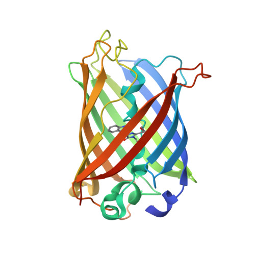
Chromophore Isomer Stabilization Is Critical to the Efficient Fluorescence of Cyan Fluorescent Proteins.

Gotthard, G.von Stetten, D.Clavel, D.Noirclerc-Savoye, M.Royant, A.

(2017) Biochemistry 56: 6418-6422

  • DOI: https://doi.org/10.1021/acs.biochem.7b01088
  • Primary Citation Related Structures: 
    5OX8, 5OX9, 5OXA, 5OXB, 5OXC

  • PubMed Abstract: 

    ECFP, the first usable cyan fluorescent protein (CFP), was obtained by adapting the tyrosine-based chromophore environment in green fluorescent protein to that of a tryptophan-based one. This first-generation CFP was superseded by the popular Cerulean, CyPet, and SCFP3A that were engineered by rational and random mutagenesis, yet the latter CFPs still exhibit suboptimal properties of pH sensitivity and reversible photobleaching behavior. These flaws were serendipitously corrected in the third-generation CFP mTurquoise and its successors without an obvious rationale. We show here that the evolution process had unexpectedly remodeled the chromophore environment in second-generation CFPs so they would accommodate a different isomer, whose formation is favored by acidic pH or light irradiation and which emits fluorescence much less efficiently. Our results illustrate how fluorescent protein engineering based solely on fluorescence efficiency optimization may affect other photophysical or physicochemical parameters and provide novel insights into the rational evolution of fluorescent proteins with a tryptophan-based chromophore.


  • Organizational Affiliation
    • European Synchrotron Radiation Facility , F-38043 Grenoble, France.

Macromolecule Content 

  • Total Structure Weight: 26.96 kDa 
  • Atom Count: 2,377 
  • Modeled Residue Count: 225 
  • Deposited Residue Count: 237 
  • Unique protein chains: 1

Macromolecules

Find similar proteins by:|  3D Structure
Entity ID: 1
MoleculeChains  Sequence LengthOrganismDetailsImage
Green fluorescent protein237Aequorea victoriaMutation(s): 4 
Gene Names: GFP
UniProt
Find proteins for P42212 (Aequorea victoria)
Explore P42212 
Go to UniProtKB:  P42212
Entity Groups
Sequence Clusters30% Identity50% Identity70% Identity90% Identity95% Identity100% Identity
UniProt GroupP42212
Sequence Annotations
Expand
Reference Sequence

Small Molecules

Ligands 1 Unique
IDChains Name / Formula / InChI Key2D Diagram3D Interactions
CL

Query on CL


Download:Ideal Coordinates CCD File
B [auth A]CHLORIDE ION
Cl
VEXZGXHMUGYJMC-UHFFFAOYSA-M
Modified Residues  1 Unique
IDChains TypeFormula2D DiagramParent
B2H
Query on B2H
A
L-PEPTIDE LINKINGC17 H20 N4 O4THR, TRP, GLY

Experimental Data & Validation

Experimental Data

  • Method: X-RAY DIFFRACTION
  • Resolution: 1.29 Å
  • R-Value Free:  0.166 (Depositor), 0.166 (DCC) 
  • R-Value Work:  0.132 (Depositor), 0.130 (DCC) 
  • R-Value Observed: 0.134 (Depositor) 
Space Group: P 21 21 21
Unit Cell:
Length ( Å )Angle ( ˚ )
a = 51.247α = 90
b = 62.624β = 90
c = 69.108γ = 90
Software Package:
Software NamePurpose
REFMACrefinement
XDSdata reduction
SCALAdata scaling
PHASERphasing

Structure Validation

View Full Validation Report


Entry History 

Deposition Data

Revision History  (Full details and data files)

  • Version 1.0: 2017-11-29
    Type: Initial release
  • Version 1.1: 2017-12-06
    Changes: Database references
  • Version 1.2: 2017-12-20
    Changes: Database references
  • Version 1.3: 2024-01-17
    Changes: Data collection, Database references, Derived calculations, Refinement description
  • Version 1.4: 2024-11-06
    Changes: Structure summary