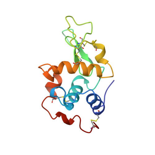
5OCV

A Rare Lysozyme Crystal Form Solved Using High-Redundancy 3D Electron Diffraction Data from Micron-Sized Needle Shaped Crystals


Experimental Data Snapshot

  • Method: ELECTRON CRYSTALLOGRAPHY
  • Resolution: 2.20 Å
  • R-Value Free: 0.270 
  • R-Value Work: 0.236 
  • R-Value Observed: 0.238 

wwPDB Validation   3D Report Full Report


This is version 1.2 of the entry. See complete history


Literature

A Rare Lysozyme Crystal Form Solved Using Highly Redundant Multiple Electron Diffraction Datasets from Micron-Sized Crystals.

Xu, H.Lebrette, H.Yang, T.Srinivas, V.Hovmoller, S.Hogbom, M.Zou, X.

(2018) Structure 26: 667-675.e3

  • DOI: https://doi.org/10.1016/j.str.2018.02.015
  • Primary Citation of Related Structures:  
    5OCV

  • PubMed Abstract: 

    Recent developments of novel electron diffraction techniques have shown to be powerful for determination of atomic resolution structures from micron- and nano-sized crystals, too small to be studied by single-crystal X-ray diffraction. In this work, the structure of a rare lysozyme polymorph is solved and refined using continuous rotation MicroED data and standard X-ray crystallographic software. Data collection was performed on a standard 200 kV transmission electron microscope (TEM) using a highly sensitive detector with a short readout time. The data collection is fast (∼3 min per crystal), allowing multiple datasets to be rapidly collected from a large number of crystals. We show that merging data from 33 crystals significantly improves not only the data completeness, overall I/σ and the data redundancy, but also the quality of the final atomic model. This is extremely useful for electron beam-sensitive crystals of low symmetry or with a preferred orientation on the TEM grid.


  • Organizational Affiliation

    Inorganic and Structural Chemistry, Department of Materials and Environmental Chemistry, Stockholm University, 106 91 Stockholm, Sweden.


Macromolecules
Find similar proteins by:  (by identity cutoff)  |  3D Structure
Entity ID: 1
MoleculeChains Sequence LengthOrganismDetailsImage
Lysozyme C
A, B
129Gallus gallusMutation(s): 0 
EC: 3.2.1.17
UniProt
Find proteins for P00698 (Gallus gallus)
Explore P00698 
Go to UniProtKB:  P00698
Entity Groups  
Sequence Clusters30% Identity50% Identity70% Identity90% Identity95% Identity100% Identity
UniProt GroupP00698
Sequence Annotations
Expand
  • Reference Sequence
Small Molecules
Ligands 1 Unique
IDChains Name / Formula / InChI Key2D Diagram3D Interactions
NA (Subject of Investigation/LOI)
Query on NA

Download Ideal Coordinates CCD File 
C [auth B]SODIUM ION
Na
FKNQFGJONOIPTF-UHFFFAOYSA-N
Experimental Data & Validation

Experimental Data

  • Method: ELECTRON CRYSTALLOGRAPHY
  • Resolution: 2.20 Å
  • R-Value Free: 0.270 
  • R-Value Work: 0.236 
  • R-Value Observed: 0.238 
  • Space Group: P 21 21 2
Unit Cell:
Length ( Å )Angle ( ˚ )
a = 67.72α = 90
b = 103.88β = 90
c = 31.65γ = 90
Software Package:
Software NamePurpose
PHENIXrefinement
XDSdata reduction
PHASERphasing
EM Software:
TaskSoftware PackageVersion
MODEL REFINEMENTPHENIX1.11.1
MODEL REFINEMENTCoot0.8.8

Structure Validation

View Full Validation Report


Entry History & Funding Information

Deposition Data


Funding OrganizationLocationGrant Number
Knut and Alice Wallenberg FoundationSweden3DEM-NATUR
Knut and Alice Wallenberg FoundationSwedenWallenberg Academy Fellows
Science for Life LaboratorySwedenElectron Nanocrystallography
Swedish Foundation for Strategic ResearchSweden--
Swedish Research CouncilSweden--
Wenner-Gren FoundationSweden--

Revision History  (Full details and data files)

  • Version 1.0: 2018-03-28
    Type: Initial release
  • Version 1.1: 2018-04-11
    Changes: Data collection, Database references
  • Version 1.2: 2024-01-17
    Changes: Data collection, Database references, Derived calculations, Refinement description