Experimental Data Snapshot

  • Method: X-RAY DIFFRACTION
  • Resolution: 1.70 Å
  • R-Value Free: 
    0.184 (Depositor), 0.178 (DCC) 
  • R-Value Work: 
    0.151 (Depositor), 0.151 (DCC) 
  • R-Value Observed: 
    0.153 (Depositor) 

Starting Model: experimental
View more details

wwPDB Validation 3D Report Full Report

Validation slider image for 5O8A

This is version 1.6 of the entry. See complete history

Literature

Chromophore twisting in the excited state of a photoswitchable fluorescent protein captured by time-resolved serial femtosecond crystallography.

Coquelle, N.Sliwa, M.Woodhouse, J.Schiro, G.Adam, V.Aquila, A.Barends, T.R.M.Boutet, S.Byrdin, M.Carbajo, S.De la Mora, E.Doak, R.B.Feliks, M.Fieschi, F.Foucar, L.Guillon, V.Hilpert, M.Hunter, M.S.Jakobs, S.Koglin, J.E.Kovacsova, G.Lane, T.J.Levy, B.Liang, M.Nass, K.Ridard, J.Robinson, J.S.Roome, C.M.Ruckebusch, C.Seaberg, M.Thepaut, M.Cammarata, M.Demachy, I.Field, M.Shoeman, R.L.Bourgeois, D.Colletier, J.P.Schlichting, I.Weik, M.

(2018) Nat Chem 10: 31-37

  • DOI: https://doi.org/10.1038/nchem.2853
  • Primary Citation Related Structures: 
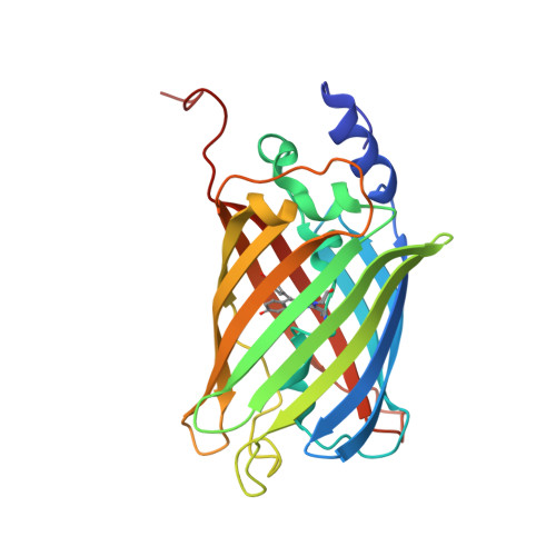
    5O89, 5O8A, 5O8B, 5O8C

  • PubMed Abstract: 

    Chromophores absorb light in photosensitive proteins and thereby initiate fundamental biological processes such as photosynthesis, vision and biofluorescence. An important goal in their understanding is the provision of detailed structural descriptions of the ultrafast photochemical events that they undergo, in particular of the excited states that connect chemistry to biological function. Here we report on the structures of two excited states in the reversibly photoswitchable fluorescent protein rsEGFP2. We populated the states through femtosecond illumination of rsEGFP2 in its non-fluorescent off state and observed their build-up (within less than one picosecond) and decay (on the several picosecond timescale). Using an X-ray free-electron laser, we performed picosecond time-resolved crystallography and show that the hydroxybenzylidene imidazolinone chromophore in one of the excited states assumes a near-canonical twisted configuration halfway between the trans and cis isomers. This is in line with excited-state quantum mechanics/molecular mechanics and classical molecular dynamics simulations. Our new understanding of the structure around the twisted chromophore enabled the design of a mutant that displays a twofold increase in its off-to-on photoswitching quantum yield.


  • Organizational Affiliation
    • Institut de Biologie Structurale, Univ. Grenoble Alpes, CEA, CNRS, F-38044 Grenoble, France.

Macromolecule Content 

  • Total Structure Weight: 28.1 kDa 
  • Atom Count: 4,233 
  • Modeled Residue Count: 244 
  • Deposited Residue Count: 246 
  • Unique protein chains: 1

Macromolecules

Find similar proteins by:|  3D Structure
Entity ID: 1
MoleculeChains  Sequence LengthOrganismDetailsImage
Green fluorescent protein246Aequorea victoriaMutation(s): 6 
Gene Names: GFP
UniProt
Find proteins for A0A1S5SHT2 (Human respiratory syncytial virus)
Explore A0A1S5SHT2 
Go to UniProtKB:  A0A1S5SHT2
Entity Groups
Sequence Clusters30% Identity50% Identity70% Identity90% Identity95% Identity100% Identity
UniProt GroupA0A1S5SHT2
Sequence Annotations
Expand
Reference Sequence

Small Molecules

Modified Residues  1 Unique
IDChains TypeFormula2D DiagramParent
PIA
Query on PIA
A
L-PEPTIDE LINKINGC14 H15 N3 O4ALA, TYR, GLY

Experimental Data & Validation

Experimental Data

  • Method: X-RAY DIFFRACTION
  • Resolution: 1.70 Å
  • R-Value Free:  0.184 (Depositor), 0.178 (DCC) 
  • R-Value Work:  0.151 (Depositor), 0.151 (DCC) 
  • R-Value Observed: 0.153 (Depositor) 
Space Group: P 21 21 21
Unit Cell:
Length ( Å )Angle ( ˚ )
a = 51.53α = 90
b = 62.85β = 90
c = 71.76γ = 90
Software Package:
Software NamePurpose
PHENIXrefinement
CrystFELdata reduction
CrystFELdata scaling
PHASERphasing

Structure Validation

View Full Validation Report


Entry History 

Revision History  (Full details and data files)

  • Version 1.0: 2017-09-27
    Type: Initial release
  • Version 1.1: 2017-12-13
    Changes: Data collection, Database references, Source and taxonomy, Structure summary
  • Version 1.2: 2017-12-27
    Changes: Database references
  • Version 1.3: 2018-11-14
    Changes: Data collection
  • Version 1.4: 2019-10-16
    Changes: Data collection
  • Version 1.5: 2023-12-13
    Changes: Advisory, Data collection, Database references, Derived calculations
  • Version 1.6: 2024-01-17
    Changes: Refinement description