Protein structure determination by electron diffraction using a single three-dimensional nanocrystal.
Clabbers, M.T.B., van Genderen, E., Wan, W., Wiegers, E.L., Gruene, T., Abrahams, J.P.(2017) Acta Crystallogr D Struct Biol 73: 738-748
- PubMed: 28876237 Search on PubMedSearch on PubMed Central
- DOI: https://doi.org/10.1107/S2059798317010348
- Primary Citation Related Structures:
5O4W, 5O4X - PubMed Abstract:
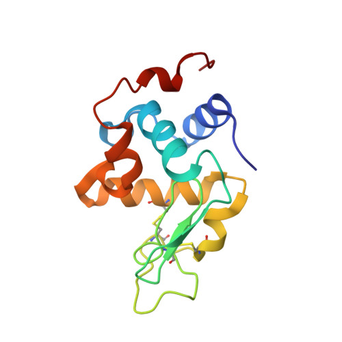
Three-dimensional nanometre-sized crystals of macromolecules currently resist structure elucidation by single-crystal X-ray crystallography. Here, a single nanocrystal with a diffracting volume of only 0.14 µm 3 , i.e. no more than 6 × 10 5 unit cells, provided sufficient information to determine the structure of a rare dimeric polymorph of hen egg-white lysozyme by electron crystallography. This is at least an order of magnitude smaller than was previously possible. The molecular-replacement solution, based on a monomeric polyalanine model, provided sufficient phasing power to show side-chain density, and automated model building was used to reconstruct the side chains. Diffraction data were acquired using the rotation method with parallel beam diffraction on a Titan Krios transmission electron microscope equipped with a novel in-house-designed 1024 × 1024 pixel Timepix hybrid pixel detector for low-dose diffraction data collection. Favourable detector characteristics include the ability to accurately discriminate single high-energy electrons from X-rays and count them, fast readout to finely sample reciprocal space and a high dynamic range. This work, together with other recent milestones, suggests that electron crystallography can provide an attractive alternative in determining biological structures.
- Center for Cellular Imaging and NanoAnalytics (C-CINA), Biozentrum, Basel University, Mattenstrasse 26, CH-4058 Basel, Switzerland.
Organizational Affiliation: