5O0K | pdb_00005o0k

Deglycosylated Nogo Receptor with native disulfide structure


Experimental Data Snapshot

  • Method: X-RAY DIFFRACTION
  • Resolution: 2.30 Å
  • R-Value Free: 
    0.234 (Depositor), 0.219 (DCC) 
  • R-Value Work: 
    0.179 (Depositor) 
  • R-Value Observed: 
    0.181 (Depositor) 

Starting Model: experimental
View more details

wwPDB Validation 3D Report Full Report

Validation slider image for 5O0K

Ligand Structure Quality Assessment 


This is version 1.5 of the entry. See complete history

Literature

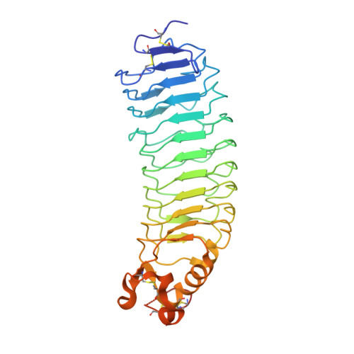
Nogo Receptor crystal structures with a native disulfide pattern suggest a novel mode of self-interaction.

Pronker, M.F.Tas, R.P.Vlieg, H.C.Janssen, B.J.C.

(2017) Acta Crystallogr D Struct Biol 73: 860-876

  • DOI: https://doi.org/10.1107/S2059798317013791
  • Primary Citation Related Structures: 
    5O0K, 5O0L, 5O0M, 5O0N, 5O0O, 5O0P, 5O0Q, 5O0R

  • PubMed Abstract: 

    The Nogo Receptor (NgR) is a glycophosphatidylinositol-anchored cell-surface protein and is a receptor for three myelin-associated inhibitors of regeneration: myelin-associated glycoprotein, Nogo66 and oligodendrocyte myelin glycoprotein. In combination with different co-receptors, NgR mediates signalling that reduces neuronal plasticity. The available structures of the NgR ligand-binding leucine-rich repeat (LRR) domain have an artificial disulfide pattern owing to truncated C-terminal construct boundaries. NgR has previously been shown to self-associate via its LRR domain, but the structural basis of this interaction remains elusive. Here, crystal structures of the NgR LRR with a longer C-terminal segment and a native disulfide pattern are presented. An additional C-terminal loop proximal to the C-terminal LRR cap is stabilized by two newly formed disulfide bonds, but is otherwise mostly unstructured in the absence of any stabilizing interactions. NgR crystallized in six unique crystal forms, three of which share a crystal-packing interface. NgR crystal-packing interfaces from all eight unique crystal forms are compared in order to explore how NgR could self-interact on the neuronal plasma membrane.


  • Organizational Affiliation
    • Crystal and Structural Chemistry, Bijvoet Center for Biomolecular Research, Department of Chemistry, Faculty of Science, Utrecht University, Padualaan 8, 3584 CH Utrecht, The Netherlands.

Macromolecule Content 

  • Total Structure Weight: 74.1 kDa 
  • Atom Count: 4,942 
  • Modeled Residue Count: 580 
  • Deposited Residue Count: 646 
  • Unique protein chains: 1

Macromolecules

Find similar proteins by:|  3D Structure
Entity ID: 1
MoleculeChains  Sequence LengthOrganismDetailsImage
Reticulon-4 receptor
A, B
323Mus musculusMutation(s): 0 
Gene Names: Rtn4rNgr1Nogor
UniProt
Find proteins for Q99PI8 (Mus musculus)
Explore Q99PI8 
Go to UniProtKB:  Q99PI8
Entity Groups
Sequence Clusters30% Identity50% Identity70% Identity90% Identity95% Identity100% Identity
UniProt GroupQ99PI8
Glycosylation
Glycosylation Sites: 3Go to GlyGen: Q99PI8-1
Sequence Annotations
Expand
Reference Sequence

Small Molecules

Ligands 3 Unique
IDChains Name / Formula / InChI Key2D Diagram3D Interactions
NAG

Query on NAG


Download:Ideal Coordinates CCD File
C [auth A]
D [auth A]
E [auth A]
K [auth B]
L [auth B]
C [auth A],
D [auth A],
E [auth A],
K [auth B],
L [auth B],
M [auth B]
2-acetamido-2-deoxy-beta-D-glucopyranose
C8 H15 N O6
OVRNDRQMDRJTHS-FMDGEEDCSA-N
GOL

Query on GOL


Download:Ideal Coordinates CCD File
F [auth A]
G [auth A]
H [auth A]
N [auth B]
O [auth B]
F [auth A],
G [auth A],
H [auth A],
N [auth B],
O [auth B],
P [auth B],
Q [auth B]
GLYCEROL
C3 H8 O3
PEDCQBHIVMGVHV-UHFFFAOYSA-N
CL

Query on CL


Download:Ideal Coordinates CCD File
I [auth A],
J [auth A],
R [auth B]
CHLORIDE ION
Cl
VEXZGXHMUGYJMC-UHFFFAOYSA-M

Experimental Data & Validation

Experimental Data

  • Method: X-RAY DIFFRACTION
  • Resolution: 2.30 Å
  • R-Value Free:  0.234 (Depositor), 0.219 (DCC) 
  • R-Value Work:  0.179 (Depositor) 
  • R-Value Observed: 0.181 (Depositor) 
Space Group: C 1 2 1
Unit Cell:
Length ( Å )Angle ( ˚ )
a = 151.624α = 90
b = 46.586β = 123.45
c = 120.67γ = 90
Software Package:
Software NamePurpose
PHENIXrefinement
XDSdata reduction
XDSdata scaling
PHASERphasing

Structure Validation

View Full Validation Report


Ligand Structure Quality Assessment 


Entry History 

& Funding Information

Deposition Data


Funding OrganizationLocationGrant Number
NWONetherlandsVidi 723.012.002

Revision History  (Full details and data files)

  • Version 1.0: 2017-10-18
    Type: Initial release
  • Version 1.1: 2017-11-08
    Changes: Database references
  • Version 1.2: 2017-11-15
    Changes: Database references
  • Version 1.3: 2020-07-29
    Type: Remediation
    Reason: Carbohydrate remediation
    Changes: Data collection, Derived calculations, Structure summary
  • Version 1.4: 2024-01-17
    Changes: Data collection, Database references, Refinement description, Structure summary
  • Version 1.5: 2024-11-13
    Changes: Structure summary