5NRO | pdb_00005nro

Structure of full-length DnaK with bound J-domain


Experimental Data Snapshot

  • Method: X-RAY DIFFRACTION
  • Resolution: 3.25 Å
  • R-Value Free: 
    0.234 (Depositor), 0.240 (DCC) 
  • R-Value Work: 
    0.216 (Depositor), 0.220 (DCC) 
  • R-Value Observed: 
    0.217 (Depositor) 

Starting Model: experimental
View more details

wwPDB Validation   3D Report Full Report


Ligand Structure Quality Assessment 

Created with Raphaël 2.3.0Worse 01 BetterLigand structure goodness of fit to experimental dataBest fitted ATPClick on this verticalbar to view details

This is version 1.3 of the entry. See complete history


Literature

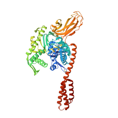
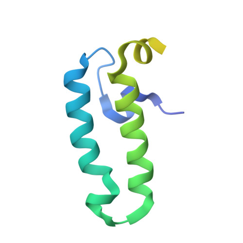
Molecular Mechanism of J-Domain-Triggered ATP Hydrolysis by Hsp70 Chaperones.

Kityk, R.Kopp, J.Mayer, M.P.

(2018) Mol Cell 69: 227-237.e4

  • DOI: https://doi.org/10.1016/j.molcel.2017.12.003
  • Primary Citation of Related Structures:  
    5NRO

  • PubMed Abstract: 

    Efficient targeting of Hsp70 chaperones to substrate proteins depends on J-domain cochaperones, which in synergism with substrates trigger ATP hydrolysis in Hsp70s and concomitant substrate trapping. We present the crystal structure of the J-domain of Escherichia coli DnaJ in complex with the E. coli Hsp70 DnaK. The J-domain interacts not only with DnaK's nucleotide-binding domain (NBD) but also with its substrate-binding domain (SBD) and packs against the highly conserved interdomain linker. Mutational replacement of contacts between J-domain and SBD strongly reduces the ability of substrates to stimulate ATP hydrolysis in the presence of DnaJ and compromises viability at heat shock temperatures. Our data demonstrate that the J-domain and the substrate do not deliver completely independent signals for ATP hydrolysis, but the J-domain, in addition to its direct influence on Hsp70s catalytic center, makes Hsp70 more responsive for the hydrolysis-inducing signal of the substrate, resulting in efficient substrate trapping.


  • Organizational Affiliation

    Center for Molecular Biology of Heidelberg University (ZMBH), DKFZ-ZMBH Alliance, 69120 Heidelberg, Germany.


Macromolecules
Find similar proteins by:  (by identity cutoff)  |  3D Structure
Entity ID: 1
MoleculeChains Sequence LengthOrganismDetailsImage
Chaperone protein DnaK611Escherichia coliMutation(s): 3 
Gene Names: dnaKgroPgrpFsegb0014JW0013
UniProt
Find proteins for P0A6Y8 (Escherichia coli (strain K12))
Explore P0A6Y8 
Go to UniProtKB:  P0A6Y8
Entity Groups  
Sequence Clusters30% Identity50% Identity70% Identity90% Identity95% Identity100% Identity
UniProt GroupP0A6Y8
Sequence Annotations
Expand
  • Reference Sequence
Find similar proteins by:  (by identity cutoff)  |  3D Structure
Entity ID: 2
MoleculeChains Sequence LengthOrganismDetailsImage
Chaperone protein DnaJ108Escherichia coliMutation(s): 0 
Gene Names: dnaJgroPb0015JW0014
UniProt
Find proteins for P08622 (Escherichia coli (strain K12))
Explore P08622 
Go to UniProtKB:  P08622
Entity Groups  
Sequence Clusters30% Identity50% Identity70% Identity90% Identity95% Identity100% Identity
UniProt GroupP08622
Sequence Annotations
Expand
  • Reference Sequence
Experimental Data & Validation

Experimental Data

  • Method: X-RAY DIFFRACTION
  • Resolution: 3.25 Å
  • R-Value Free:  0.234 (Depositor), 0.240 (DCC) 
  • R-Value Work:  0.216 (Depositor), 0.220 (DCC) 
  • R-Value Observed: 0.217 (Depositor) 
Space Group: P 62 2 2
Unit Cell:
Length ( Å )Angle ( ˚ )
a = 127.246α = 90
b = 127.246β = 90
c = 252.885γ = 120
Software Package:
Software NamePurpose
PHENIXrefinement
XDSdata reduction
Aimlessdata scaling
PHASERphasing

Structure Validation

View Full Validation Report


Ligand Structure Quality Assessment 

Created with Raphaël 2.3.0Worse 01 BetterLigand structure goodness of fit to experimental dataBest fitted ATPClick on this verticalbar to view details

Entry History & Funding Information

Deposition Data


Funding OrganizationLocationGrant Number
German Research FoundationGermanyMA 1278/4-3

Revision History  (Full details and data files)

  • Version 1.0: 2018-01-17
    Type: Initial release
  • Version 1.1: 2018-01-31
    Changes: Database references
  • Version 1.2: 2024-01-17
    Changes: Data collection, Database references, Refinement description
  • Version 1.3: 2024-11-06
    Changes: Structure summary