Structural insights into the tyrosine phosphorylation-mediated inhibition of SH3 domain-ligand interactions.
Mero, B., Radnai, L., Gogl, G., Toke, O., Leveles, I., Koprivanacz, K., Szeder, B., Dulk, M., Kudlik, G., Vas, V., Cserkaszky, A., Sipeki, S., Nyitray, L., Vertessy, B.G., Buday, L.(2019) J Biological Chem 294: 4608-4620
- PubMed: 30659095 Search on PubMedSearch on PubMed Central
- DOI: https://doi.org/10.1074/jbc.RA118.004732
- Primary Citation Related Structures:
5NP2, 5NP3, 5NP5 - PubMed Abstract:
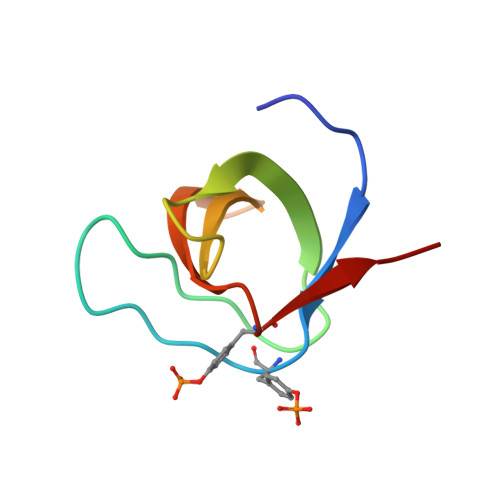
Src homology 3 (SH3) domains bind proline-rich linear motifs in eukaryotes. By mediating inter- and intramolecular interactions, they regulate the functions of many proteins involved in a wide variety of signal transduction pathways. Phosphorylation at different tyrosine residues in SH3 domains has been reported previously. In several cases, the functional consequences have also been investigated. However, a full understanding of the effects of tyrosine phosphorylation on the ligand interactions and cellular functions of SH3 domains requires detailed structural, atomic-resolution studies along with biochemical and biophysical analyses. Here, we present the first crystal structures of tyrosine-phosphorylated human SH3 domains derived from the Abelson-family kinases ABL1 and ABL2 at 1.6 and 1.4 Å resolutions, respectively. The structures revealed that simultaneous phosphorylation of Tyr 89 and Tyr 134 in ABL1 or the homologous residues Tyr 116 and Tyr 161 in ABL2 induces only minor structural perturbations. Instead, the phosphate groups sterically blocked the ligand-binding grooves, thereby strongly inhibiting the interaction with proline-rich peptide ligands. Although some crystal contact surfaces involving phosphotyrosines suggested the possibility of tyrosine phosphorylation-induced dimerization, we excluded this possibility by using small-angle X-ray scattering (SAXS), dynamic light scattering (DLS), and NMR relaxation analyses. Extensive analysis of relevant databases and literature revealed not only that the residues phosphorylated in our model systems are well-conserved in other human SH3 domains, but that the corresponding tyrosines are known phosphorylation sites in vivo in many cases. We conclude that tyrosine phosphorylation might be a mechanism involved in the regulation of the human SH3 interactome.
- From the Institute of Enzymology and.
Organizational Affiliation: