Aggregation Pathways of Native-Like Ubiquitin Promoted by Single-Point Mutation, Metal Ion Concentration, and Dielectric Constant of the Medium.
Fermani, S., Calvaresi, M., Mangini, V., Falini, G., Bottoni, A., Natile, G., Arnesano, F.(2018) Chemistry 24: 4140-4148
- PubMed: 29266436 Search on PubMed
- DOI: https://doi.org/10.1002/chem.201705543
- Primary Citation Related Structures:
5NL4, 5NL5, 5NLF, 5NLI, 5NLJ, 5NMC - PubMed Abstract:
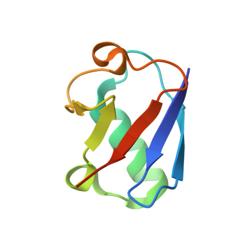
Ubiquitin-positive protein aggregates are biomarkers of neurodegeneration, but the molecular mechanism responsible for their formation and accumulation is still unclear. Possible aggregation pathways of human ubiquitin (hUb) promoted by both intrinsic and extrinsic factors, are here investigated. By a computational analysis, two different hUb dimers are indicated as possible precursors of amyloid-like structures, but their formation is disfavored by an electrostatic repulsion involving Glu16 and other carboxylate residues present at the dimer interface. Experimental data on the E16V mutant of hUb shows that this single-point mutation, although not affecting the overall protein conformation, promotes protein aggregation. It is sufficient to shift the same mutation by only two residues (E18V) to regain the behavior of wild-type hUb. The neutralization of Glu16 negative charge by a metal ion and a decrease of the dielectric constant of the medium by addition of trifluoroethanol (TFE), also promote hUb aggregation. The outcomes of this research have important implications for the prediction of physiological parameters that favor aggregate formation.
- Department of Chemistry "Giacomo Ciamician", University of Bologna, via F. Selmi 2, 40126, Bologna, Italy.
Organizational Affiliation: