5NIJ | pdb_00005nij

Crystal structure of arabidopsis thaliana legumain isoform gamma in two-chain activation state


Experimental Data Snapshot

  • Method: X-RAY DIFFRACTION
  • Resolution: 2.75 Å
  • R-Value Free: 
    0.252 (Depositor), 0.253 (DCC) 
  • R-Value Work: 
    0.214 (Depositor), 0.222 (DCC) 
  • R-Value Observed: 
    0.215 (Depositor) 

Starting Model: experimental
View more details

wwPDB Validation 3D Report Full Report

Validation slider image for 5NIJ

This is version 2.1 of the entry. See complete history

Literature

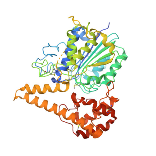
Crystal Structure of Plant Legumain Reveals a Unique Two-Chain State with pH-Dependent Activity Regulation.

Zauner, F.B.Dall, E.Regl, C.Grassi, L.Huber, C.G.Cabrele, C.Brandstetter, H.

(2018) Plant Cell 30: 686-699

  • DOI: https://doi.org/10.1105/tpc.17.00963
  • Primary Citation Related Structures: 
    5NIJ

  • PubMed Abstract: 

    The vacuolar cysteine protease legumain can cleave and selectively rebuild peptide bonds, thereby vastly expanding the sequential repertoire of biomolecules. In this context, plant legumains have recently attracted particular interest. Furthermore, legumains have important roles in many physiological processes, including programmed cell death. Their efficient peptide bond ligase activity has gained tremendous interest in the design of cyclic peptides for drug design. However, the mechanistic understanding of these dual activities is incomplete and partly conflicting. Here, we present the crystal structure of a plant legumain, Arabidopsis thaliana isoform-γ (AtLEGγ). Employing a conserved legumain fold, the plant legumain AtLEGγ revealed unique mechanisms of autoactivation, including a plant-specific two-chain activation state, which remains conformationally stable at neutral pH, which is a prerequisite for full ligase activity and survival in different cell compartments. The charge distribution around the α6-helix mediates the pH-dependent dimerization and serves as a gatekeeper for the active site, thus regulating its protease and ligase activity.


  • Organizational Affiliation
    • Department of Molecular Biology and Christian Doppler Laboratory for Biosimilar Research, University of Salzburg, A-5020 Salzburg, Austria.

Macromolecule Content 

  • Total Structure Weight: 202.81 kDa 
  • Atom Count: 13,474 
  • Modeled Residue Count: 1,714 
  • Deposited Residue Count: 1,816 
  • Unique protein chains: 1

Macromolecules

Find similar proteins by:|  3D Structure
Entity ID: 1
MoleculeChains  Sequence LengthOrganismDetailsImage
Vacuolar-processing enzyme gamma-isozyme
A, B, C, D
454Arabidopsis thalianaMutation(s): 0 
Gene Names: At4g32940F26P21.60
EC: 3.4.22.34
UniProt
Find proteins for Q39119 (Arabidopsis thaliana)
Explore Q39119 
Go to UniProtKB:  Q39119
Entity Groups
Sequence Clusters30% Identity50% Identity70% Identity90% Identity95% Identity100% Identity
UniProt GroupQ39119
Sequence Annotations
Expand
Reference Sequence

Experimental Data & Validation

Experimental Data

  • Method: X-RAY DIFFRACTION
  • Resolution: 2.75 Å
  • R-Value Free:  0.252 (Depositor), 0.253 (DCC) 
  • R-Value Work:  0.214 (Depositor), 0.222 (DCC) 
  • R-Value Observed: 0.215 (Depositor) 
Space Group: P 41
Unit Cell:
Length ( Å )Angle ( ˚ )
a = 147.83α = 90
b = 147.83β = 90
c = 101.6γ = 90
Software Package:
Software NamePurpose
PHENIXrefinement
iMOSFLMdata reduction
Aimlessdata scaling
PHASERphasing

Structure Validation

View Full Validation Report


Entry History 

& Funding Information

Deposition Data


Funding OrganizationLocationGrant Number
Austrian Science FundAustriaW_01213

Revision History  (Full details and data files)

  • Version 1.0: 2018-02-28
    Type: Initial release
  • Version 1.1: 2018-04-18
    Changes: Data collection, Database references
  • Version 2.0: 2023-11-15
    Changes: Atomic model, Data collection, Database references, Derived calculations
  • Version 2.1: 2024-01-17
    Changes: Refinement description