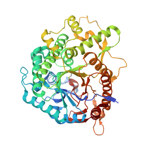
Structural basis of the substrate specificity and instability in solution of a glycosidase from Lactobacillus plantarum.
Acebron, I., Plaza-Vinuesa, L., de Las Rivas, B., Munoz, R., Cumella, J., Sanchez-Sancho, F., Mancheno, J.M.(2017) Biochim Biophys Acta 1865: 1227-1236
- PubMed: 28734976
- DOI: https://doi.org/10.1016/j.bbapap.2017.07.007
- Primary Citation of Related Structures:
5NAQ, 5NAV - PubMed Abstract:
Statistics from structural genomics initiatives reveal that around 50-55% of the expressed, non-membrane proteins cannot be purified and therefore structurally characterized due to solubility problems, which emphasized protein solubility as one of the most serious concerns in structural biology projects. Lactobacillus plantarum CECT 748 T produces an aggregation-prone glycosidase (LpBgl) that we crystallized previously. However, this result could not be reproduced due to protein instability and therefore further high-resolution structural analyses of LpBgl were impeded. The obtained crystals of LpBgl diffracted up to 2.48Å resolution and permitted to solve the structure of the enzyme. Analysis of the active site revealed a pocket for phosphate-binding with an uncommon architecture, where a phosphate molecule is tightly bound suggesting the recognition of 6-phosphoryl sugars. In agreement with this observation, we showed that LpBgl exhibited 6-phospho-β-glucosidase activity. Combination of structural and mass spectrometry results revealed the formation of dimethyl arsenic adducts on the solvent exposed cysteine residues Cys211 and Cys292. Remarkably, the double mutant Cys211Ser/Cys292Ser resulted stable in solution at high concentrations indicating that the marginal solubility of LpBgl can be ascribed specifically to these two cysteine residues. The 2.30Å crystal structure of this double mutant showed no disorder around the newly incorporated serine residues and also loop rearrangements within the phosphate-binding site. Notably, LpBgl could be prepared at high yield by proteolytic digestion of the fusion protein LSLt-LpBgl, which raises important questions about potential hysteretic processes upon its initial production as an enzyme fused to a solubility enhancer.
- Department of Crystallography and Structural Biology, Institute of Physical Chemistry Rocasolano, CSIC, Serrano 119, E-28006 Madrid, Spain.
Organizational Affiliation:


