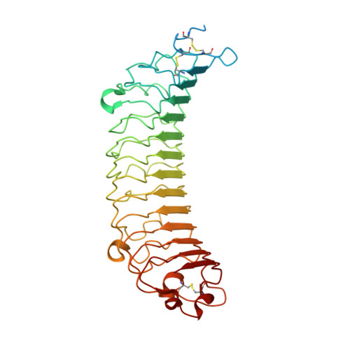
5MX0

Crystal structure of human fibromodulin


Experimental Data Snapshot

  • Method: X-RAY DIFFRACTION
  • Resolution: 2.21 Å
  • R-Value Free: 0.216 
  • R-Value Work: 0.179 
  • R-Value Observed: 0.181 

wwPDB Validation   3D Report Full Report


This is version 2.1 of the entry. See complete history


Literature

Structural and functional analysis of two small leucine-rich repeat proteoglycans, fibromodulin and chondroadherin.

Paracuellos, P.Kalamajski, S.Bonna, A.Bihan, D.Farndale, R.W.Hohenester, E.

(2017) Matrix Biol 63: 106-116

  • DOI: https://doi.org/10.1016/j.matbio.2017.02.002
  • Primary Citation of Related Structures:  
    5MX0, 5MX1

  • PubMed Abstract: 

    The small leucine-rich proteoglycans (SLRPs) are important regulators of extracellular matrix assembly and cell signalling. We have determined crystal structures at ~2.2Å resolution of human fibromodulin and chondroadherin, two collagen-binding SLRPs. Their overall fold is similar to that of the prototypical SLRP, decorin, but unlike decorin neither fibromodulin nor chondroadherin forms a stable dimer. A previously identified binding site for integrin α2β1 maps to an α-helix in the C-terminal cap region of chondroadherin. Interrogation of the Collagen Toolkits revealed a unique binding site for chondroadherin in collagen II, and no binding to collagen III. A triple-helical peptide containing the sequence GAOGPSGFQGLOGPOGPO (O is hydroxyproline) forms a stable complex with chondroadherin in solution. In fibrillar collagen I and II, this sequence is aligned with the collagen cross-linking site KGHR, suggesting a role for chondroadherin in cross-linking.


  • Organizational Affiliation

    Department of Life Sciences, Imperial College London, London, United Kingdom.


Macromolecules
Find similar proteins by:  (by identity cutoff)  |  3D Structure
Entity ID: 1
MoleculeChains Sequence LengthOrganismDetailsImage
Fibromodulin
A, B
362Homo sapiensMutation(s): 10 
Gene Names: FMODFMSLRR2E
UniProt & NIH Common Fund Data Resources
Find proteins for Q06828 (Homo sapiens)
Explore Q06828 
Go to UniProtKB:  Q06828
PHAROS:  Q06828
GTEx:  ENSG00000122176 
Entity Groups  
Sequence Clusters30% Identity50% Identity70% Identity90% Identity95% Identity100% Identity
UniProt GroupQ06828
Sequence Annotations
Expand
  • Reference Sequence
Oligosaccharides

Help

Entity ID: 2
MoleculeChains Length2D Diagram Glycosylation3D Interactions
beta-D-mannopyranose-(1-4)-2-acetamido-2-deoxy-beta-D-glucopyranose-(1-4)-[alpha-L-fucopyranose-(1-6)]2-acetamido-2-deoxy-beta-D-glucopyranose
C, D, E, F, G
C, D, E, F, G, H, I, J
4N-Glycosylation
Glycosylation Resources
GlyTouCan:  G32152BH
GlyCosmos:  G32152BH
GlyGen:  G32152BH
Experimental Data & Validation

Experimental Data

  • Method: X-RAY DIFFRACTION
  • Resolution: 2.21 Å
  • R-Value Free: 0.216 
  • R-Value Work: 0.179 
  • R-Value Observed: 0.181 
  • Space Group: C 1 2 1
Unit Cell:
Length ( Å )Angle ( ˚ )
a = 108.11α = 90
b = 98.93β = 107.39
c = 111.34γ = 90
Software Package:
Software NamePurpose
PHENIXrefinement
xia2data reduction
xia2data scaling
PHENIXphasing

Structure Validation

View Full Validation Report


Entry History & Funding Information

Deposition Data


Funding OrganizationLocationGrant Number
Wellcome TrustUnited Kingdom101748/Z/13/Z

Revision History  (Full details and data files)

  • Version 1.0: 2017-03-01
    Type: Initial release
  • Version 1.1: 2017-09-27
    Changes: Database references
  • Version 2.0: 2020-07-29
    Type: Remediation
    Reason: Carbohydrate remediation
    Changes: Atomic model, Data collection, Derived calculations, Structure summary
  • Version 2.1: 2024-01-17
    Changes: Data collection, Database references, Derived calculations, Refinement description, Structure summary