5MSD

Structure of the A domain of carboxylic acid reductase (CAR) from Nocardia iowensis in complex with AMP and benzoic acid


Experimental Data Snapshot

  • Method: X-RAY DIFFRACTION
  • Resolution: 1.71 Å
  • R-Value Free: 0.215 
  • R-Value Work: 0.180 
  • R-Value Observed: 0.182 

wwPDB Validation   3D Report Full Report


Ligand Structure Quality Assessment 


This is version 1.5 of the entry. See complete history


Literature

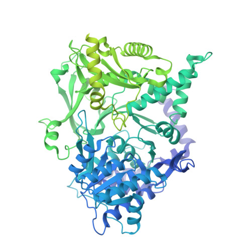
Structures of carboxylic acid reductase reveal domain dynamics underlying catalysis.

Gahloth, D.Dunstan, M.S.Quaglia, D.Klumbys, E.Lockhart-Cairns, M.P.Hill, A.M.Derrington, S.R.Scrutton, N.S.Turner, N.J.Leys, D.

(2017) Nat Chem Biol 13: 975-981

  • DOI: https://doi.org/10.1038/nchembio.2434
  • Primary Citation of Related Structures:  
    5MSC, 5MSD, 5MSO, 5MSP, 5MSR, 5MSS, 5MST, 5MSU, 5MSV, 5MSW

  • PubMed Abstract: 

    Carboxylic acid reductase (CAR) catalyzes the ATP- and NADPH-dependent reduction of carboxylic acids to the corresponding aldehydes. The enzyme is related to the nonribosomal peptide synthetases, consisting of an adenylation domain fused via a peptidyl carrier protein (PCP) to a reductase termination domain. Crystal structures of the CAR adenylation-PCP didomain demonstrate that large-scale domain motions occur between the adenylation and thiolation states. Crystal structures of the PCP-reductase didomain reveal that phosphopantetheine binding alters the orientation of a key Asp, resulting in a productive orientation of the bound nicotinamide. This ensures that further reduction of the aldehyde product does not occur. Combining crystallography with small-angle X-ray scattering (SAXS), we propose that molecular interactions between initiation and termination domains are limited to competing PCP docking sites. This theory is supported by the fact that (R)-pantetheine can support CAR activity for mixtures of the isolated domains. Our model suggests directions for further development of CAR as a biocatalyst.


  • Organizational Affiliation

    Manchester Institute of Biotechnology, School of Chemistry, University of Manchester, Manchester, UK.


Macromolecules
Find similar proteins by:  (by identity cutoff)  |  3D Structure
Entity ID: 1
MoleculeChains Sequence LengthOrganismDetailsImage
Carboxylic acid reductase1,174Nocardia iowensisMutation(s): 0 
Gene Names: car
EC: 1.2.1 (PDB Primary Data), 1.2.1.30 (PDB Primary Data)
UniProt
Find proteins for Q6RKB1 (Nocardia iowensis)
Explore Q6RKB1 
Go to UniProtKB:  Q6RKB1
Entity Groups  
Sequence Clusters30% Identity50% Identity70% Identity90% Identity95% Identity100% Identity
UniProt GroupQ6RKB1
Sequence Annotations
Expand
  • Reference Sequence
Small Molecules
Ligands 2 Unique
IDChains Name / Formula / InChI Key2D Diagram3D Interactions
AMP
Query on AMP

Download Ideal Coordinates CCD File 
B [auth A]ADENOSINE MONOPHOSPHATE
C10 H14 N5 O7 P
UDMBCSSLTHHNCD-KQYNXXCUSA-N
BEZ
Query on BEZ

Download Ideal Coordinates CCD File 
C [auth A]BENZOIC ACID
C7 H6 O2
WPYMKLBDIGXBTP-UHFFFAOYSA-N
Experimental Data & Validation

Experimental Data

  • Method: X-RAY DIFFRACTION
  • Resolution: 1.71 Å
  • R-Value Free: 0.215 
  • R-Value Work: 0.180 
  • R-Value Observed: 0.182 
  • Space Group: P 1
Unit Cell:
Length ( Å )Angle ( ˚ )
a = 52.65α = 93.37
b = 54.499β = 97.68
c = 66.365γ = 102.44
Software Package:
Software NamePurpose
PHENIXrefinement
XDSdata reduction
XDSdata scaling

Structure Validation

View Full Validation Report


Ligand Structure Quality Assessment 


Entry History 

Deposition Data

Revision History  (Full details and data files)

  • Version 1.0: 2017-07-05
    Type: Initial release
  • Version 1.1: 2017-07-12
    Changes: Database references
  • Version 1.2: 2017-07-26
    Changes: Database references
  • Version 1.3: 2017-08-30
    Changes: Database references
  • Version 1.4: 2019-10-16
    Changes: Data collection
  • Version 1.5: 2024-01-17
    Changes: Data collection, Database references, Refinement description