5MNJ

Structure of MDM2-MDMX-UbcH5B-ubiquitin complex


Experimental Data Snapshot

  • Method: X-RAY DIFFRACTION
  • Resolution: 2.16 Å
  • R-Value Free: 0.231 
  • R-Value Work: 0.190 
  • R-Value Observed: 0.192 

wwPDB Validation   3D Report Full Report


This is version 1.5 of the entry. See complete history


Literature

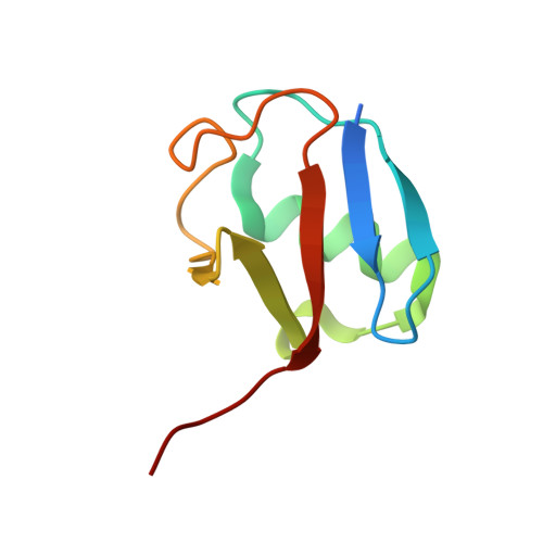
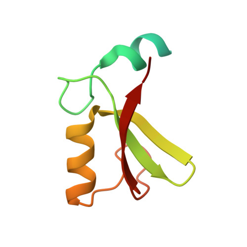
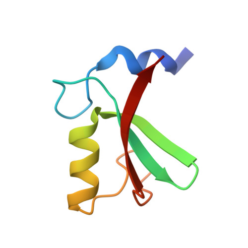
Structural analysis of MDM2 RING separates degradation from regulation of p53 transcription activity.

Nomura, K.Klejnot, M.Kowalczyk, D.Hock, A.K.Sibbet, G.J.Vousden, K.H.Huang, D.T.

(2017) Nat Struct Mol Biol 24: 578-587

  • DOI: 10.1038/nsmb.3414
  • Primary Citation of Related Structures:  
    5MNJ

  • PubMed Abstract: 
  • MDM2-MDMX complexes bind the p53 tumor-suppressor protein, inhibiting p53's transcriptional activity and targeting p53 for proteasomal degradation. Inhibitors that disrupt binding between p53 and MDM2 efficiently activate a p53 response, but their use in the treatment of cancers that retain wild-type p53 may be limited by on-target toxicities due to p53 activation in normal tissue ...

    MDM2-MDMX complexes bind the p53 tumor-suppressor protein, inhibiting p53's transcriptional activity and targeting p53 for proteasomal degradation. Inhibitors that disrupt binding between p53 and MDM2 efficiently activate a p53 response, but their use in the treatment of cancers that retain wild-type p53 may be limited by on-target toxicities due to p53 activation in normal tissue. Guided by a novel crystal structure of the MDM2-MDMX-E2(UbcH5B)-ubiquitin complex, we designed MDM2 mutants that prevent E2-ubiquitin binding without altering the RING-domain structure. These mutants lack MDM2's E3 activity but retain the ability to limit p53's transcriptional activity and allow cell proliferation. Cells expressing these mutants respond more quickly to cellular stress than cells expressing wild-type MDM2, but basal p53 control is maintained. Targeting the MDM2 E3-ligase activity could therefore widen the therapeutic window of p53 activation in tumors.


    Organizational Affiliation

    Cancer Research-UK Beatson Institute, Garscube Estate, Glasgow, UK.


Macromolecules
Find similar proteins by:  (by identity cutoff)  |  3D Structure
Entity ID: 1
MoleculeChainsSequence LengthOrganismDetailsImage
Ubiquitin-conjugating enzyme E2 D2A, E147Homo sapiensMutation(s): 2 
Gene Names: UBE2D2PUBC1UBC4UBC5BUBCH4UBCH5B
EC: 2.3.2.23 (PDB Primary Data), 2.3.2.24 (PDB Primary Data)
UniProt & NIH Common Fund Data Resources
Find proteins for P62837 (Homo sapiens)
Explore P62837 
Go to UniProtKB:  P62837
PHAROS:  P62837
Entity Groups  
Sequence Clusters30% Identity50% Identity70% Identity90% Identity95% Identity100% Identity
UniProt GroupP62837
Protein Feature View
Expand
  • Reference Sequence
Find similar proteins by:  (by identity cutoff)  |  3D Structure
Entity ID: 2
MoleculeChainsSequence LengthOrganismDetailsImage
Polyubiquitin-BB, F81Homo sapiensMutation(s): 0 
Gene Names: UBB
UniProt & NIH Common Fund Data Resources
Find proteins for P0CG47 (Homo sapiens)
Explore P0CG47 
Go to UniProtKB:  P0CG47
PHAROS:  P0CG47
Entity Groups  
Sequence Clusters30% Identity50% Identity70% Identity90% Identity95% Identity100% Identity
UniProt GroupP0CG47
Protein Feature View
Expand
  • Reference Sequence
Find similar proteins by:  (by identity cutoff)  |  3D Structure
Entity ID: 3
MoleculeChainsSequence LengthOrganismDetailsImage
E3 ubiquitin-protein ligase Mdm2C, G86Homo sapiensMutation(s): 0 
Gene Names: MDM2
EC: 6.3.2 (PDB Primary Data), 2.3.2.27 (UniProt)
UniProt & NIH Common Fund Data Resources
Find proteins for Q00987 (Homo sapiens)
Explore Q00987 
Go to UniProtKB:  Q00987
PHAROS:  Q00987
Entity Groups  
Sequence Clusters30% Identity50% Identity70% Identity90% Identity95% Identity100% Identity
UniProt GroupQ00987
Protein Feature View
Expand
  • Reference Sequence
Find similar proteins by:  (by identity cutoff)  |  3D Structure
Entity ID: 4
MoleculeChainsSequence LengthOrganismDetailsImage
Protein Mdm4D, H64Homo sapiensMutation(s): 0 
Gene Names: MDM4MDMX
UniProt & NIH Common Fund Data Resources
Find proteins for O15151 (Homo sapiens)
Explore O15151 
Go to UniProtKB:  O15151
PHAROS:  O15151
Entity Groups  
Sequence Clusters30% Identity50% Identity70% Identity90% Identity95% Identity100% Identity
UniProt GroupO15151
Protein Feature View
Expand
  • Reference Sequence
Experimental Data & Validation

Experimental Data

  • Method: X-RAY DIFFRACTION
  • Resolution: 2.16 Å
  • R-Value Free: 0.231 
  • R-Value Work: 0.190 
  • R-Value Observed: 0.192 
  • Space Group: P 1
Unit Cell:
Length ( Å )Angle ( ˚ )
a = 54.24α = 69.83
b = 62.76β = 69.22
c = 66.35γ = 78.21
Software Package:
Software NamePurpose
PHENIXrefinement
XDSdata reduction
xia2data scaling
PHASERphasing

Structure Validation

View Full Validation Report


Entry History & Funding Information

Deposition Data


Funding OrganizationLocationGrant Number
Cancer Research UKUnited Kingdom--
European Research Council647849

Revision History  (Full details and data files)

  • Version 1.0: 2017-05-31
    Type: Initial release
  • Version 1.1: 2017-06-07
    Changes: Database references
  • Version 1.2: 2017-07-19
    Changes: Database references
  • Version 1.3: 2019-05-08
    Changes: Advisory, Data collection, Derived calculations
  • Version 1.4: 2019-07-10
    Changes: Data collection
  • Version 1.5: 2019-10-16
    Changes: Data collection