5MME

Crystal structure of CREBBP bromodomain complexd with US46C


Experimental Data Snapshot

  • Method: X-RAY DIFFRACTION
  • Resolution: 1.35 Å
  • R-Value Free: 0.176 
  • R-Value Work: 0.164 
  • R-Value Observed: 0.164 

wwPDB Validation   3D Report Full Report


Ligand Structure Quality Assessment 


This is version 1.3 of the entry. See complete history


Literature

Binding Motifs in the CBP Bromodomain: An Analysis of 20 Crystal Structures of Complexes with Small Molecules.

Zhu, J.Dong, J.Batiste, L.Unzue, A.Dolbois, A.Pascanu, V.Sledz, P.Nevado, C.Caflisch, A.

(2018) ACS Med Chem Lett 9: 929-934

  • DOI: https://doi.org/10.1021/acsmedchemlett.8b00286
  • Primary Citation of Related Structures:  
    5EIC, 5ENG, 5EP7, 5H85, 5MME, 5MMG, 5MPK, 5MPN, 5OWK, 6FQO, 6FQU, 6FR0, 6FRF

  • PubMed Abstract: 

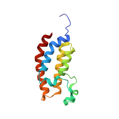
    We analyze 20 crystal structures of complexes between the CBP bromodomain and small-molecule ligands that belong to eight different chemotypes identified by docking. The binding motif of the moiety that mimics the natural ligand (acetylated side chain of lysine) at the bottom of the binding pocket is conserved. In stark contrast, the rest of the ligands form different interactions with different side chains and backbone polar groups on the outer rim of the binding pocket. Hydrogen bonds are direct or water-bridged. van der Waals contacts are optimized by rotations of hydrophobic side chains and a slight inward displacement of the ZA loop. Rare types of interactions are observed for some of the ligands.


  • Organizational Affiliation

    Department of Biochemistry, and Department of Chemistry, University of Zurich, Winterthurerstrasse 190, CH-8057 Zurich, Switzerland.


Macromolecules
Find similar proteins by:  (by identity cutoff)  |  3D Structure
Entity ID: 1
MoleculeChains Sequence LengthOrganismDetailsImage
CREB-binding protein
A, B
119Homo sapiensMutation(s): 0 
Gene Names: CREBBPCBP
EC: 2.3.1.48
UniProt & NIH Common Fund Data Resources
Find proteins for Q92793 (Homo sapiens)
Explore Q92793 
Go to UniProtKB:  Q92793
PHAROS:  Q92793
GTEx:  ENSG00000005339 
Entity Groups  
Sequence Clusters30% Identity50% Identity70% Identity90% Identity95% Identity100% Identity
UniProt GroupQ92793
Sequence Annotations
Expand
  • Reference Sequence
Small Molecules
Ligands 1 Unique
IDChains Name / Formula / InChI Key2D Diagram3D Interactions
8Q6
Query on 8Q6

Download Ideal Coordinates CCD File 
C [auth A],
D [auth B]
dimethyl 5-[(5-ethanoyl-2-ethoxy-phenyl)amino]benzene-1,3-dicarboxylate
C20 H21 N O6
UNAGVZCODUSBMG-UHFFFAOYSA-N
Binding Affinity Annotations 
IDSourceBinding Affinity
8Q6 BindingDB:  5MME Kd: 1400 (nM) from 1 assay(s)
Binding MOAD:  5MME Kd: 1400 (nM) from 1 assay(s)
Experimental Data & Validation

Experimental Data

  • Method: X-RAY DIFFRACTION
  • Resolution: 1.35 Å
  • R-Value Free: 0.176 
  • R-Value Work: 0.164 
  • R-Value Observed: 0.164 
  • Space Group: P 21 21 21
Unit Cell:
Length ( Å )Angle ( ˚ )
a = 53.371α = 90
b = 54.265β = 90
c = 81.048γ = 90
Software Package:
Software NamePurpose
PHENIXrefinement
XDSdata reduction
XDSdata scaling
PHASERphasing

Structure Validation

View Full Validation Report


Ligand Structure Quality Assessment 


Entry History 

Deposition Data

Revision History  (Full details and data files)

  • Version 1.0: 2017-12-20
    Type: Initial release
  • Version 1.1: 2018-10-10
    Changes: Data collection, Database references
  • Version 1.2: 2019-10-16
    Changes: Data collection
  • Version 1.3: 2024-01-17
    Changes: Data collection, Database references, Refinement description