5MK8

Crystal structure of the receptor-binding domain of the FA hybrid Clostridium botulinum neurotoxin


Experimental Data Snapshot

  • Method: X-RAY DIFFRACTION
  • Resolution: 1.95 Å
  • R-Value Free: 0.212 
  • R-Value Work: 0.179 
  • R-Value Observed: 0.181 

wwPDB Validation   3D Report Full Report


This is version 1.1 of the entry. See complete history


Literature

High resolution crystal structures of the receptor-binding domain ofClostridium botulinumneurotoxin serotypes A and FA.

Davies, J.R.Hackett, G.S.Liu, S.M.Acharya, K.R.

(2018) PeerJ 6: e4552-e4552

  • DOI: https://doi.org/10.7717/peerj.4552
  • Primary Citation of Related Structures:  
    5MK6, 5MK7, 5MK8

  • PubMed Abstract: 

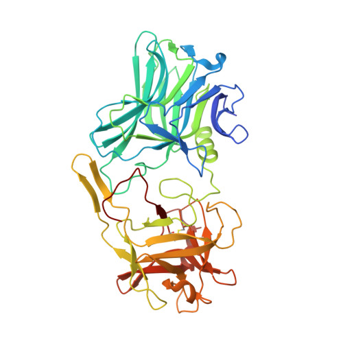
    The binding specificity of botulinum neurotoxins (BoNTs) is primarily a consequence of their ability to bind to multiple receptors at the same time. BoNTs consist of three distinct domains, a metalloprotease light chain (LC), a translocation domain (H N ) and a receptor-binding domain (H C ). Here we report the crystal structure of H C /FA, complementing an existing structure through the modelling of a previously unresolved loop which is important for receptor-binding. Our H C /FA structure also contains a previously unidentified disulphide bond, which we have also observed in one of two crystal forms of H C /A1. This may have implications for receptor-binding and future recombinant toxin production.


  • Organizational Affiliation

    Department of Biology and Biochemistry, University of Bath, Bath, United Kingdom.


Macromolecules
Find similar proteins by:  (by identity cutoff)  |  3D Structure
Entity ID: 1
MoleculeChains Sequence LengthOrganismDetailsImage
Botulinum neurotoxin FA binding domain
A, B
437Clostridium botulinumMutation(s): 0 
UniProt
Find proteins for Q9R540 (Clostridium botulinum)
Explore Q9R540 
Go to UniProtKB:  Q9R540
Entity Groups  
Sequence Clusters30% Identity50% Identity70% Identity90% Identity95% Identity100% Identity
UniProt GroupQ9R540
Sequence Annotations
Expand
  • Reference Sequence
Small Molecules
Ligands 2 Unique
IDChains Name / Formula / InChI Key2D Diagram3D Interactions
FMT
Query on FMT

Download Ideal Coordinates CCD File 
C [auth A]
D [auth A]
E [auth A]
F [auth A]
G [auth A]
C [auth A],
D [auth A],
E [auth A],
F [auth A],
G [auth A],
H [auth A],
K [auth B],
L [auth B],
M [auth B],
N [auth B],
O [auth B]
FORMIC ACID
C H2 O2
BDAGIHXWWSANSR-UHFFFAOYSA-N
CL
Query on CL

Download Ideal Coordinates CCD File 
I [auth A],
J [auth A],
P [auth B],
Q [auth B]
CHLORIDE ION
Cl
VEXZGXHMUGYJMC-UHFFFAOYSA-M
Experimental Data & Validation

Experimental Data

  • Method: X-RAY DIFFRACTION
  • Resolution: 1.95 Å
  • R-Value Free: 0.212 
  • R-Value Work: 0.179 
  • R-Value Observed: 0.181 
  • Space Group: P 4 2 2
Unit Cell:
Length ( Å )Angle ( ˚ )
a = 118.009α = 90
b = 118.009β = 90
c = 173.826γ = 90
Software Package:
Software NamePurpose
PHENIXrefinement
DIALSdata reduction
Aimlessdata scaling
PHASERphasing

Structure Validation

View Full Validation Report


Entry History 

Deposition Data

Revision History  (Full details and data files)

  • Version 1.0: 2018-03-28
    Type: Initial release
  • Version 1.1: 2018-04-04
    Changes: Data collection, Database references