5MJA | pdb_00005mja

Kinase domain of human EphB1 bound to a quinazoline-based inhibitor


Experimental Data Snapshot

  • Method: X-RAY DIFFRACTION
  • Resolution: 2.14 Å
  • R-Value Free: 
    0.236 (Depositor), 0.220 (DCC) 
  • R-Value Work: 
    0.200 (Depositor), 0.200 (DCC) 
  • R-Value Observed: 
    0.202 (Depositor) 

Starting Model: experimental
View more details

wwPDB Validation   3D Report Full Report


Ligand Structure Quality Assessment 

Created with Raphaël 2.3.0Worse 01 BetterLigand structure goodness of fit to experimental dataBest fitted 7O3Click on this verticalbar to view details

This is version 1.4 of the entry. See complete history


Literature

A Chemical-Genetic Approach to Generate Selective Covalent Inhibitors of Protein Kinases.

Kung, A.Schimpl, M.Ekanayake, A.Chen, Y.C.Overman, R.Zhang, C.

(2017) ACS Chem Biol 12: 1499-1503

  • DOI: https://doi.org/10.1021/acschembio.6b01083
  • Primary Citation of Related Structures:  
    5MJA, 5MJB

  • PubMed Abstract: 

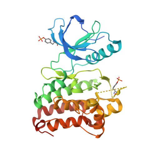
    Although a previously developed bump-hole approach has proven powerful in generating specific inhibitors for mapping functions of protein kinases, its application is limited by the intolerance of the large-to-small mutation by certain kinases and the inability to control two kinases separately in the same cells. Herein, we describe the development of an alternative chemical-genetic approach to overcome these limitations. Our approach features the use of an engineered cysteine residue at a particular position as a reactive feature to sensitize a kinase of interest to selective covalent blockade by electrophilic inhibitors and is thus termed the Ele-Cys approach. We successfully applied the Ele-Cys approach to identify selective covalent inhibitors of a receptor tyrosine kinase EphB1 and solved cocrystal structures to determine the mode of covalent binding. Importantly, the Ele-Cys and bump-hole approaches afforded orthogonal inhibition of two distinct kinases in the cell, opening the door to their combined use in the study of multikinase signaling pathways.


  • Organizational Affiliation

    Discovery Sciences, Innovative Medicines and Early Development Biotech Unit, AstraZeneca , Building 310, Cambridge Science Park, Milton Road, Cambridge, CB4 0WG, United Kingdom.


Macromolecules
Find similar proteins by:  (by identity cutoff)  |  3D Structure
Entity ID: 1
MoleculeChains Sequence LengthOrganismDetailsImage
Ephrin type-B receptor 1
A, B
305Homo sapiensMutation(s): 0 
Gene Names: EPHB1ELKEPHT2HEK6NET
EC: 2.7.10.1
UniProt & NIH Common Fund Data Resources
Find proteins for P54762 (Homo sapiens)
Explore P54762 
Go to UniProtKB:  P54762
PHAROS:  P54762
GTEx:  ENSG00000154928 
Entity Groups  
Sequence Clusters30% Identity50% Identity70% Identity90% Identity95% Identity100% Identity
UniProt GroupP54762
Sequence Annotations
Expand
  • Reference Sequence
Experimental Data & Validation

Experimental Data

  • Method: X-RAY DIFFRACTION
  • Resolution: 2.14 Å
  • R-Value Free:  0.236 (Depositor), 0.220 (DCC) 
  • R-Value Work:  0.200 (Depositor), 0.200 (DCC) 
  • R-Value Observed: 0.202 (Depositor) 
Space Group: P 31 2 1
Unit Cell:
Length ( Å )Angle ( ˚ )
a = 101.806α = 90
b = 101.806β = 90
c = 157.14γ = 120
Software Package:
Software NamePurpose
BUSTERrefinement
DIALSdata reduction
Aimlessdata scaling
BUSTERphasing

Structure Validation

View Full Validation Report


Ligand Structure Quality Assessment 

Created with Raphaël 2.3.0Worse 01 BetterLigand structure goodness of fit to experimental dataBest fitted 7O3Click on this verticalbar to view details

Entry History 

Deposition Data

Revision History  (Full details and data files)

  • Version 1.0: 2017-05-17
    Type: Initial release
  • Version 1.1: 2017-06-28
    Changes: Database references
  • Version 1.2: 2018-01-24
    Changes: Source and taxonomy
  • Version 1.3: 2024-01-17
    Changes: Data collection, Database references, Refinement description
  • Version 1.4: 2024-11-20
    Changes: Structure summary