5MHH | pdb_00005mhh

Crystal structure of engineered human lipocalin 2 carrying p-boronophenylalanine at position 36


Experimental Data Snapshot

  • Method: X-RAY DIFFRACTION
  • Resolution: 2.00 Å
  • R-Value Free: 
    0.233 (Depositor), 0.239 (DCC) 
  • R-Value Work: 
    0.181 (Depositor), 0.189 (DCC) 
  • R-Value Observed: 
    0.184 (Depositor) 

Starting Model: experimental
View more details

wwPDB Validation 3D Report Full Report

Validation slider image for 5MHH

This is version 2.2 of the entry. See complete history

Literature

Rational Design of an Anticalin-Type Sugar-Binding Protein Using a Genetically Encoded Boronate Side Chain.

Edwardraja, S.Eichinger, A.Theobald, I.Sommer, C.A.Reichert, A.J.Skerra, A.

(2017) ACS Synth Biol 6: 2241-2247

  • DOI: https://doi.org/10.1021/acssynbio.7b00199
  • Primary Citation Related Structures: 
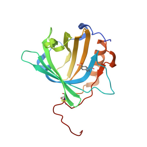
    5MHH

  • PubMed Abstract: 

    The molecular recognition of carbohydrates plays a fundamental role in many biological processes. However, the development of carbohydrate-binding reagents for biomedical research and use poses a challenge due to the generally poor affinity of proteins toward sugars in aqueous solution. Here, we describe the effective molecular recognition of pyranose monosaccharides (in particular, galactose and mannose) by a rationally designed protein receptor based on the human lipocalin scaffold (Anticalin). Complexation relies on reversible covalent cis-diol boronate diester formation with a genetically encoded l-boronophenylalanine (Bpa) residue which was incorporated as a non-natural amino acid at a sterically permissive position in the ligand pocket of the Anticalin, as confirmed by X-ray crystallography. Compared with the metal-ion and/or avidity-dependent oligovalent lectins that prevail in nature, our approach offers a novel and promising route to generate tight sugar-binding reagents both as research reagents and for biomedical applications.


  • Organizational Affiliation
    • Munich Center for Integrated Protein Science (CIPS-M) and Lehrstuhl für Biologische Chemie, Technische Universität München , Emil-Erlenmeyer-Forum 5, 85354 Freising (Weihenstephan), Germany.

Macromolecule Content 

  • Total Structure Weight: 22.16 kDa 
  • Atom Count: 1,557 
  • Modeled Residue Count: 182 
  • Deposited Residue Count: 188 
  • Unique protein chains: 1

Macromolecules

Find similar proteins by:|  3D Structure
Entity ID: 1
MoleculeChains  Sequence LengthOrganismDetailsImage
Neutrophil gelatinase-associated lipocalin188Homo sapiensMutation(s): 5 
Gene Names: LCN2HNLNGAL
UniProt & NIH Common Fund Data Resources
Find proteins for P80188 (Homo sapiens)
Explore P80188 
Go to UniProtKB:  P80188
PHAROS:  P80188
GTEx:  ENSG00000148346 
Entity Groups
Sequence Clusters30% Identity50% Identity70% Identity90% Identity95% Identity100% Identity
UniProt GroupP80188
Sequence Annotations
Expand
Reference Sequence

Experimental Data & Validation

Experimental Data

  • Method: X-RAY DIFFRACTION
  • Resolution: 2.00 Å
  • R-Value Free:  0.233 (Depositor), 0.239 (DCC) 
  • R-Value Work:  0.181 (Depositor), 0.189 (DCC) 
  • R-Value Observed: 0.184 (Depositor) 
Space Group: I 4
Unit Cell:
Length ( Å )Angle ( ˚ )
a = 90.857α = 90
b = 90.857β = 90
c = 44.2γ = 90
Software Package:
Software NamePurpose
MOSFLMdata reduction
SCALAdata scaling
PHASERphasing
REFMACrefinement
PDB_EXTRACTdata extraction

Structure Validation

View Full Validation Report


Entry History 

& Funding Information

Deposition Data


Funding OrganizationLocationGrant Number
European Molecular Biology OrganizationGermanyALTF number 858-2011
Helmholtz-Zentrum BerlinGermany--

Revision History  (Full details and data files)

  • Version 1.0: 2017-10-11
    Type: Initial release
  • Version 1.1: 2017-12-27
    Changes: Database references
  • Version 2.0: 2019-04-24
    Changes: Data collection, Polymer sequence
  • Version 2.1: 2024-01-17
    Changes: Data collection, Database references, Derived calculations, Refinement description
  • Version 2.2: 2024-10-16
    Changes: Structure summary