5M8J | pdb_00005m8j

Crystal structure of Eremococcus coleocola manganese transporter mutant H236A


Experimental Data Snapshot

  • Method: X-RAY DIFFRACTION
  • Resolution: 3.70 Å
  • R-Value Free: 
    0.257 (Depositor), 0.334 (DCC) 
  • R-Value Work: 
    0.210 (Depositor), 0.266 (DCC) 
  • R-Value Observed: 
    0.213 (Depositor) 

Starting Model: experimental
View more details

wwPDB Validation 3D Report Full Report

Validation slider image for 5M8J

This is version 1.3 of the entry. See complete history

Literature

Structural and mechanistic basis of proton-coupled metal ion transport in the SLC11/NRAMP family.

Ehrnstorfer, I.A.Manatschal, C.Arnold, F.M.Laederach, J.Dutzler, R.

(2017) Nat Commun 8: 14033-14033

  • DOI: https://doi.org/10.1038/ncomms14033
  • Primary Citation Related Structures: 
    5M87, 5M8A, 5M8J, 5M8K

  • PubMed Abstract: 

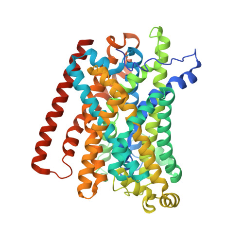
    Secondary active transporters of the SLC11/NRAMP family catalyse the uptake of iron and manganese into cells. These proteins are highly conserved across all kingdoms of life and thus likely share a common transport mechanism. Here we describe the structural and functional properties of the prokaryotic SLC11 transporter EcoDMT. Its crystal structure reveals a previously unknown outward-facing state of the protein family. In proteoliposomes EcoDMT mediates proton-coupled uptake of manganese at low micromolar concentrations. Mutants of residues in the transition-metal ion-binding site severely affect transport, whereas a mutation of a conserved histidine located near this site results in metal ion transport that appears uncoupled to proton transport. Combined with previous results, our study defines the conformational changes underlying transition-metal ion transport in the SLC11 family and it provides molecular insight to its coupling to protons.


  • Organizational Affiliation
    • Department of Biochemistry, University of Zurich, Winterthurerstrasse 190, 8057 Zurich, Switzerland.

Macromolecule Content 

  • Total Structure Weight: 56.56 kDa 
  • Atom Count: 3,775 
  • Modeled Residue Count: 494 
  • Deposited Residue Count: 519 
  • Unique protein chains: 1

Macromolecules

Find similar proteins by:|  3D Structure
Entity ID: 1
MoleculeChains  Sequence LengthOrganismDetailsImage
Divalent metal cation transporter MntH519Eremococcus coleocola ACS-139-V-Col8Mutation(s): 1 
Gene Names: mntHHMPREF9257_1603
Membrane Entity: Yes 
UniProt
Find proteins for E4KPW4 (Eremococcus coleocola ACS-139-V-Col8)
Explore E4KPW4 
Go to UniProtKB:  E4KPW4
Entity Groups
Sequence Clusters30% Identity50% Identity70% Identity90% Identity95% Identity100% Identity
UniProt GroupE4KPW4
Sequence Annotations
Expand
Reference Sequence

Experimental Data & Validation

Experimental Data

  • Method: X-RAY DIFFRACTION
  • Resolution: 3.70 Å
  • R-Value Free:  0.257 (Depositor), 0.334 (DCC) 
  • R-Value Work:  0.210 (Depositor), 0.266 (DCC) 
  • R-Value Observed: 0.213 (Depositor) 
Space Group: C 1 2 1
Unit Cell:
Length ( Å )Angle ( ˚ )
a = 150.4α = 90
b = 81.79β = 107.47
c = 96.44γ = 90
Software Package:
Software NamePurpose
XDSdata reduction
XDSdata scaling
PHENIXrefinement
PHENIXphasing

Structure Validation

View Full Validation Report


Entry History 

& Funding Information

Deposition Data


Funding OrganizationLocationGrant Number
Swiss National Science FoundationSwitzerland--

Revision History  (Full details and data files)

  • Version 1.0: 2017-01-11
    Type: Initial release
  • Version 1.1: 2017-01-18
    Changes: Database references
  • Version 1.2: 2017-09-13
    Changes: Author supporting evidence
  • Version 1.3: 2024-01-17
    Changes: Data collection, Database references, Refinement description