5M2O

R. flavefaciens' third ScaB cohesin in complex with a group 1 dockerin


Experimental Data Snapshot

  • Method: X-RAY DIFFRACTION
  • Resolution: 1.26 Å
  • R-Value Free: 0.153 
  • R-Value Work: 0.135 
  • R-Value Observed: 0.136 

Starting Model: experimental
View more details

wwPDB Validation   3D Report Full Report


This is version 1.1 of the entry. See complete history


Literature

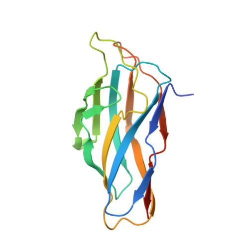
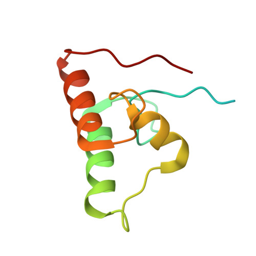
Assembly of Ruminococcus flavefaciens cellulosome revealed by structures of two cohesin-dockerin complexes.

Bule, P.Alves, V.D.Israeli-Ruimy, V.Carvalho, A.L.Ferreira, L.M.Smith, S.P.Gilbert, H.J.Najmudin, S.Bayer, E.A.Fontes, C.M.

(2017) Sci Rep 7: 759-759

  • DOI: https://doi.org/10.1038/s41598-017-00919-w
  • Primary Citation of Related Structures:  
    5M2O, 5M2S

  • PubMed Abstract: 

    Cellulosomes are sophisticated multi-enzymatic nanomachines produced by anaerobes to effectively deconstruct plant structural carbohydrates. Cellulosome assembly involves the binding of enzyme-borne dockerins (Doc) to repeated cohesin (Coh) modules located in a non-catalytic scaffoldin. Docs appended to cellulosomal enzymes generally present two similar Coh-binding interfaces supporting a dual-binding mode, which may confer increased positional adjustment of the different complex components. Ruminococcus flavefaciens' cellulosome is assembled from a repertoire of 223 Doc-containing proteins classified into 6 groups. Recent studies revealed that Docs of groups 3 and 6 are recruited to the cellulosome via a single-binding mode mechanism with an adaptor scaffoldin. To investigate the extent to which the single-binding mode contributes to the assembly of R. flavefaciens cellulosome, the structures of two group 1 Docs bound to Cohs of primary (ScaA) and adaptor (ScaB) scaffoldins were solved. The data revealed that group 1 Docs display a conserved mechanism of Coh recognition involving a single-binding mode. Therefore, in contrast to all cellulosomes described to date, the assembly of R. flavefaciens cellulosome involves single but not dual-binding mode Docs. Thus, this work reveals a novel mechanism of cellulosome assembly and challenges the ubiquitous implication of the dual-binding mode in the acquisition of cellulosome flexibility.


  • Organizational Affiliation

    CIISA - Faculdade de Medicina Veterinária, ULisboa, Pólo Universitário do Alto da Ajuda, Avenida da Universidade Técnica, 1300-477, Lisboa, Portugal.


Macromolecules
Find similar proteins by:  (by identity cutoff)  |  3D Structure
Entity ID: 1
MoleculeChains Sequence LengthOrganismDetailsImage
Putative cellulosomal scaffoldin protein149Ruminococcus flavefaciens FD-1Mutation(s): 0 
Gene Names: scaB
UniProt
Find proteins for A0AEF4 (Ruminococcus flavefaciens)
Explore A0AEF4 
Go to UniProtKB:  A0AEF4
Entity Groups  
Sequence Clusters30% Identity50% Identity70% Identity90% Identity95% Identity100% Identity
UniProt GroupA0AEF4
Sequence Annotations
Expand
  • Reference Sequence
Find similar proteins by:  (by identity cutoff)  |  3D Structure
Entity ID: 2
MoleculeChains Sequence LengthOrganismDetailsImage
Group I Dockerin96Ruminococcus flavefaciens FD-1Mutation(s): 0 
Entity Groups  
Sequence Clusters30% Identity50% Identity70% Identity90% Identity95% Identity100% Identity
Sequence Annotations
Expand
  • Reference Sequence
Experimental Data & Validation

Experimental Data

  • Method: X-RAY DIFFRACTION
  • Resolution: 1.26 Å
  • R-Value Free: 0.153 
  • R-Value Work: 0.135 
  • R-Value Observed: 0.136 
  • Space Group: P 21 21 21
Unit Cell:
Length ( Å )Angle ( ˚ )
a = 42.767α = 90
b = 63.51β = 90
c = 84.482γ = 90
Software Package:
Software NamePurpose
Aimlessdata scaling
REFMACrefinement
PDB_EXTRACTdata extraction
iMOSFLMdata reduction
PHASERphasing

Structure Validation

View Full Validation Report


Entry History & Funding Information

Deposition Data


Funding OrganizationLocationGrant Number
Fundacao para a Ciencia e TecnologiaPortugalPTDC/BIA-MIC/5947/2014
Fundacao para a Ciencia e TecnologiaPortugalSFRH/BD/86821/2012

Revision History  (Full details and data files)

  • Version 1.0: 2017-07-05
    Type: Initial release
  • Version 1.1: 2024-01-17
    Changes: Data collection, Database references, Refinement description