5M1X | pdb_00005m1x

Crystal structure of S. cerevisiae Rfa1 N-OB domain mutant (K45E)


Experimental Data Snapshot

  • Method: X-RAY DIFFRACTION
  • Resolution: 1.80 Å
  • R-Value Free: 
    0.214 (Depositor), 0.215 (DCC) 
  • R-Value Work: 
    0.164 (Depositor), 0.175 (DCC) 
  • R-Value Observed: 
    0.167 (Depositor) 

wwPDB Validation 3D Report Full Report

Validation slider image for 5M1X

This is version 1.2 of the entry. See complete history

Literature

RPA Mediates Recruitment of MRX to Forks and Double-Strand Breaks to Hold Sister Chromatids Together.

Seeber, A.Hegnauer, A.M.Hustedt, N.Deshpande, I.Poli, J.Eglinger, J.Pasero, P.Gut, H.Shinohara, M.Hopfner, K.P.Shimada, K.Gasser, S.M.

(2016) Mol Cell 64: 951-966

  • DOI: https://doi.org/10.1016/j.molcel.2016.10.032
  • Primary Citation Related Structures: 
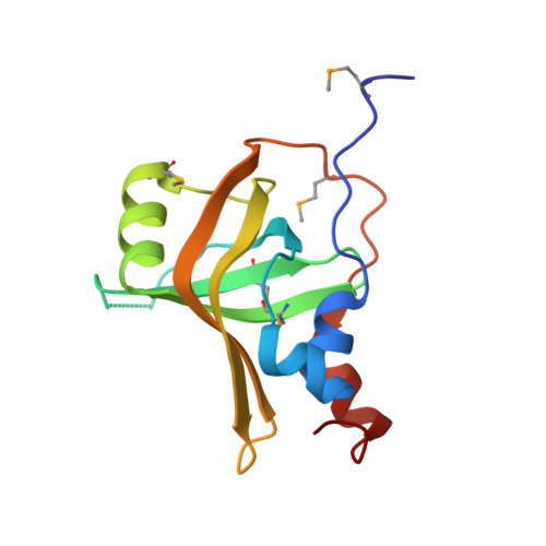
    5M1X

  • PubMed Abstract: 

    The Mre11-Rad50-Xrs2 (MRX) complex is related to SMC complexes that form rings capable of holding two distinct DNA strands together. MRX functions at stalled replication forks and double-strand breaks (DSBs). A mutation in the N-terminal OB fold of the 70 kDa subunit of yeast replication protein A, rfa1-t11, abrogates MRX recruitment to both types of DNA damage. The rfa1 mutation is functionally epistatic with loss of any of the MRX subunits for survival of replication fork stress or DSB recovery, although it does not compromise end-resection. High-resolution imaging shows that either the rfa1-t11 or the rad50Δ mutation lets stalled replication forks collapse and allows the separation not only of opposing ends but of sister chromatids at breaks. Given that cohesin loss does not provoke visible sister separation as long as the RPA-MRX contacts are intact, we conclude that MRX also serves as a structural linchpin holding sister chromatids together at breaks.


  • Organizational Affiliation
    • Friedrich Miescher Institute for Biomedical Research, Maulbeerstrasse 66, 4058 Basel, Switzerland; University of Basel, Faculty of Natural Sciences, Klingelbergstrasse 50, 4056 Basel, Switzerland.

Macromolecule Content 

  • Total Structure Weight: 62.47 kDa 
  • Atom Count: 4,653 
  • Modeled Residue Count: 518 
  • Deposited Residue Count: 536 
  • Unique protein chains: 1

Macromolecules

Find similar proteins by:|  3D Structure
Entity ID: 1
MoleculeChains  Sequence LengthOrganismDetailsImage
Replication factor A protein 1
A, B, C, D
134Saccharomyces cerevisiae S288CMutation(s): 1 
Gene Names: RFA1BUF2RPA1YAR007CFUN3
UniProt
Find proteins for P22336 (Saccharomyces cerevisiae (strain ATCC 204508 / S288c))
Explore P22336 
Go to UniProtKB:  P22336
Entity Groups
Sequence Clusters30% Identity50% Identity70% Identity90% Identity95% Identity100% Identity
UniProt GroupP22336
Sequence Annotations
Expand
Reference Sequence

Small Molecules

Modified Residues  1 Unique
IDChains TypeFormula2D DiagramParent
MSE
Query on MSE
A, B, C, D
L-PEPTIDE LINKINGC5 H11 N O2 SeMET

Experimental Data & Validation

Experimental Data

  • Method: X-RAY DIFFRACTION
  • Resolution: 1.80 Å
  • R-Value Free:  0.214 (Depositor), 0.215 (DCC) 
  • R-Value Work:  0.164 (Depositor), 0.175 (DCC) 
  • R-Value Observed: 0.167 (Depositor) 
Space Group: P 1 21 1
Unit Cell:
Length ( Å )Angle ( ˚ )
a = 29.62α = 90
b = 115.35β = 90.71
c = 69.03γ = 90
Software Package:
Software NamePurpose
BUSTERrefinement
XDSdata reduction
XSCALEdata scaling
PHENIXphasing

Structure Validation

View Full Validation Report


Entry History 

Revision History  (Full details and data files)

  • Version 1.0: 2016-12-07
    Type: Initial release
  • Version 1.1: 2016-12-14
    Changes: Database references
  • Version 1.2: 2024-10-23
    Changes: Data collection, Database references, Structure summary