


Atomic resolution X-ray crystal structure of cisplatin bound to hen egg white lysozyme stored for 5 years 'on the shelf'
Helliwell, J.R., Simon, T.(2016) Zenodo
Experimental Data Snapshot
Starting Model: experimental
View more details
wwPDB Validation 3D Report Full Report

(2016) Zenodo
Entity ID: 1 | |||||
|---|---|---|---|---|---|
| Molecule | Chains | Sequence Length | Organism | Details | Image |
| Lysozyme C | 129 | Gallus gallus | Mutation(s): 0 EC: 3.2.1.17 | 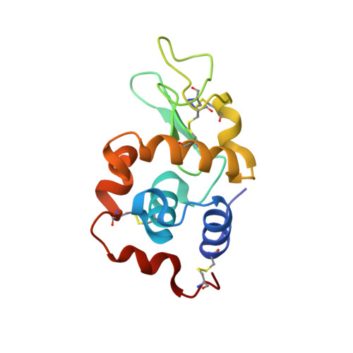 | |
UniProt | |||||
Entity Groups | |||||
| Sequence Clusters | 30% Identity50% Identity70% Identity90% Identity95% Identity100% Identity | ||||
| UniProt Group | P00698 | ||||
Sequence AnnotationsExpand | |||||
Reference Sequence | |||||
| Ligands 5 Unique | |||||
|---|---|---|---|---|---|
| ID | Chains | Name / Formula / InChI Key | 2D Diagram | 3D Interactions | |
| PT Download:Ideal Coordinates CCD File | E [auth A], F [auth A], H [auth A] | PLATINUM (II) ION Pt HRGDZIGMBDGFTC-UHFFFAOYSA-N |  | ||
| DMS Download:Ideal Coordinates CCD File | B [auth A] | DIMETHYL SULFOXIDE C2 H6 O S IAZDPXIOMUYVGZ-UHFFFAOYSA-N |  | ||
| CL Download:Ideal Coordinates CCD File | C [auth A], G [auth A], I [auth A], L [auth A], M [auth A] | CHLORIDE ION Cl VEXZGXHMUGYJMC-UHFFFAOYSA-M |  | ||
| NA Download:Ideal Coordinates CCD File | D [auth A] | SODIUM ION Na FKNQFGJONOIPTF-UHFFFAOYSA-N |  | ||
| NH3 Download:Ideal Coordinates CCD File | J [auth A], K [auth A] | AMMONIA H3 N QGZKDVFQNNGYKY-UHFFFAOYSA-N |  | ||
| Length ( Å ) | Angle ( ˚ ) |
|---|---|
| a = 79.37 | α = 90 |
| b = 79.37 | β = 90 |
| c = 37.84 | γ = 90 |
| Software Name | Purpose |
|---|---|
| PHENIX | refinement |
| iMOSFLM | data reduction |
| SCALA | data scaling |
| PHASER | phasing |


