5LSB | pdb_00005lsb

Crystal structure of yeast Hsh49p in complex with Cus1p binding domain.


Experimental Data Snapshot

  • Method: X-RAY DIFFRACTION
  • Resolution: 2.70 Å
  • R-Value Free: 
    0.233 (Depositor), 0.252 (DCC) 
  • R-Value Work: 
    0.184 (Depositor), 0.211 (DCC) 
  • R-Value Observed: 
    0.186 (Depositor) 

Starting Models: experimental
View more details

wwPDB Validation 3D Report Full Report

Validation slider image for 5LSB

This is version 1.2 of the entry. See complete history

Literature

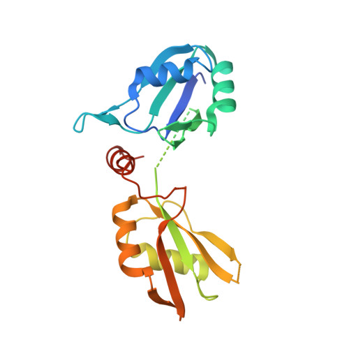
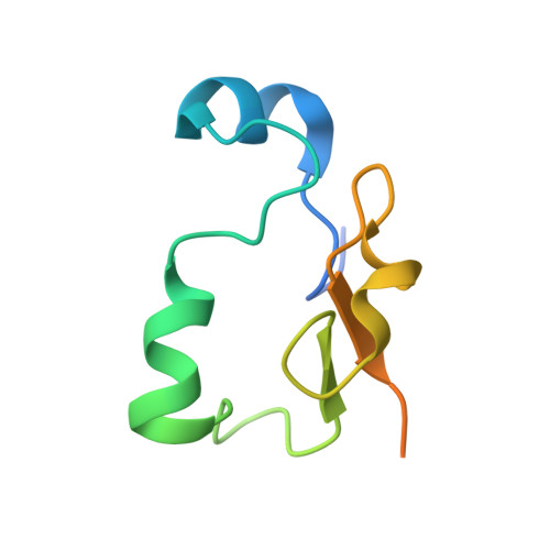
Crystal structure of U2 snRNP SF3b components: Hsh49p in complex with Cus1p-binding domain.

van Roon, A.M.Oubridge, C.Obayashi, E.Sposito, B.Newman, A.J.Seraphin, B.Nagai, K.

(2017) RNA 23: 968-981

  • DOI: https://doi.org/10.1261/rna.059378.116
  • Primary Citation Related Structures: 
    5LSB, 5LSL

  • PubMed Abstract: 

    Spliceosomal proteins Hsh49p and Cus1p are components of SF3b, which together with SF3a, Msl1p/Lea1p, Sm proteins, and U2 snRNA, form U2 snRNP, which plays a crucial role in pre-mRNA splicing. Hsh49p, comprising two RRMs, forms a heterodimer with Cus1p. We determined the crystal structures of Saccharomyces cerevisiae full-length Hsh49p as well as its RRM1 in complex with a minimal binding region of Cus1p (residues 290-368). The structures show that the Cus1 fragment binds to the α-helical surface of Hsh49p RRM1, opposite the four-stranded β-sheet, leaving the canonical RNA-binding surface available to bind RNA. Hsh49p binds the 5' end region of U2 snRNA via RRM1. Its affinity is increased in complex with Cus1(290-368)p, partly because an extended RNA-binding surface forms across the protein-protein interface. The Hsh49p RRM1-Cus1(290-368)p structure fits well into cryo-EM density of the B act spliceosome, corroborating the biological relevance of our crystal structure.


  • Organizational Affiliation
    • MRC Laboratory of Molecular Biology, Cambridge CB2 0QH, United Kingdom.

Macromolecule Content 

  • Total Structure Weight: 101.98 kDa 
  • Atom Count: 5,799 
  • Modeled Residue Count: 712 
  • Deposited Residue Count: 897 
  • Unique protein chains: 2

Macromolecules

Find similar proteins by:|  3D Structure
Entity ID: 1
MoleculeChains  Sequence LengthOrganismDetailsImage
Protein HSH49A,
C,
E [auth F]
215Saccharomyces cerevisiae S288CMutation(s): 0 
Gene Names: HSH49YOR319WO6142
UniProt
Find proteins for Q99181 (Saccharomyces cerevisiae (strain ATCC 204508 / S288c))
Explore Q99181 
Go to UniProtKB:  Q99181
Entity Groups
Sequence Clusters30% Identity50% Identity70% Identity90% Identity95% Identity100% Identity
UniProt GroupQ99181
Sequence Annotations
Expand
Reference Sequence
Find similar proteins by:|  3D Structure
Entity ID: 2
MoleculeChains  Sequence LengthOrganismDetailsImage
Cold sensitive U2 snRNA suppressor 1B,
D,
F [auth H]
84Saccharomyces cerevisiae S288CMutation(s): 0 
Gene Names: CUS1YMR240CYM9408.02C
UniProt
Find proteins for Q02554 (Saccharomyces cerevisiae (strain ATCC 204508 / S288c))
Explore Q02554 
Go to UniProtKB:  Q02554
Entity Groups
Sequence Clusters30% Identity50% Identity70% Identity90% Identity95% Identity100% Identity
UniProt GroupQ02554
Sequence Annotations
Expand
Reference Sequence

Experimental Data & Validation

Experimental Data

  • Method: X-RAY DIFFRACTION
  • Resolution: 2.70 Å
  • R-Value Free:  0.233 (Depositor), 0.252 (DCC) 
  • R-Value Work:  0.184 (Depositor), 0.211 (DCC) 
  • R-Value Observed: 0.186 (Depositor) 
Space Group: P 32
Unit Cell:
Length ( Å )Angle ( ˚ )
a = 142.083α = 90
b = 142.083β = 90
c = 40.355γ = 120
Software Package:
Software NamePurpose
REFMACrefinement
XDSdata reduction
Aimlessdata scaling
PHASERphasing
PDB_EXTRACTdata extraction

Structure Validation

View Full Validation Report


Entry History 

Deposition Data

Revision History  (Full details and data files)

  • Version 1.0: 2017-04-12
    Type: Initial release
  • Version 1.1: 2017-05-24
    Changes: Database references
  • Version 1.2: 2024-01-17
    Changes: Data collection, Database references, Refinement description