5LK4 | pdb_00005lk4

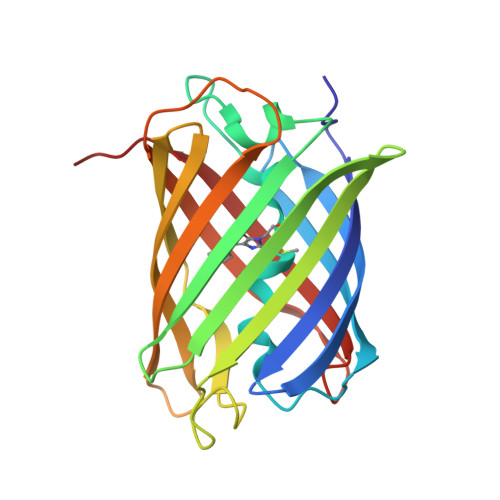
Structure of the Red Fluorescent Protein mScarlet at pH 7.8


Experimental Data Snapshot

  • Method: X-RAY DIFFRACTION
  • Resolution: 1.47 Å
  • R-Value Free: 
    0.167 (Depositor), 0.167 (DCC) 
  • R-Value Work: 
    0.137 (Depositor), 0.136 (DCC) 
  • R-Value Observed: 
    0.138 (Depositor) 

Starting Model: experimental
View more details

wwPDB Validation 3D Report Full Report

Validation slider image for 5LK4

This is version 3.0 of the entry. See complete history. 

Literature

mScarlet: a bright monomeric red fluorescent protein for cellular imaging.

Bindels, D.S., Haarbosch, L., van Weeren, L., Postma, M., Wiese, K.E., Mastop, M., Aumonier, S., Gotthard, G., Royant, A., Hink, M.A., Gadella, T.W.

(2017) Nat Methods 14: 53-56

  • DOI: https://doi.org/10.1038/nmeth.4074
  • Primary Citation Related Structures: 
    5LK4

  • PubMed Abstract: 

    We report the engineering of mScarlet, a truly monomeric red fluorescent protein with record brightness, quantum yield (70%) and fluorescence lifetime (3.9 ns). We developed mScarlet starting with a consensus synthetic template and using improved spectroscopic screening techniques; mScarlet's crystal structure reveals a planar and rigidified chromophore. mScarlet outperforms existing red fluorescent proteins as a fusion tag, and it is especially useful as a Förster resonance energy transfer (FRET) acceptor in ratiometric imaging.


  • Organizational Affiliation: 
    • Section of Molecular Cytology and van Leeuwenhoek Centre for Advanced Microscopy, Swammerdam Institute for Life Sciences, University of Amsterdam, Amsterdam, the Netherlands.

Macromolecule Content 

  • Total Structure Weight: 25.89 kDa 
  • Atom Count: 2,128 
  • Modeled Residue Count: 217 
  • Deposited Residue Count: 217 
  • Unique protein chains: 1

Macromolecules

Find similar proteins by:|  3D Structure
Entity ID: 1
MoleculeChains  Sequence LengthOrganismDetailsImage
mScarlet217Discosoma sp.Mutation(s): 40 
UniProt
Find proteins for Q9U6Y8 (Discosoma sp.)
Explore Q9U6Y8 
Go to UniProtKB:  Q9U6Y8
Entity Groups
Sequence Clusters30% Identity50% Identity70% Identity90% Identity95% Identity100% Identity
UniProt GroupQ9U6Y8
Sequence Annotations
Expand
Reference Sequence

Small Molecules

Ligands 4 Unique
IDChains Name / Formula / InChI Key2D Diagram3D Interactions
PG4

Query on PG4


Download:Ideal Coordinates CCD File
B [auth A]TETRAETHYLENE GLYCOL
C8 H18 O5
UWHCKJMYHZGTIT-UHFFFAOYSA-N
PGE

Query on PGE


Download:Ideal Coordinates CCD File
D [auth A],
E [auth A],
F [auth A],
G [auth A]
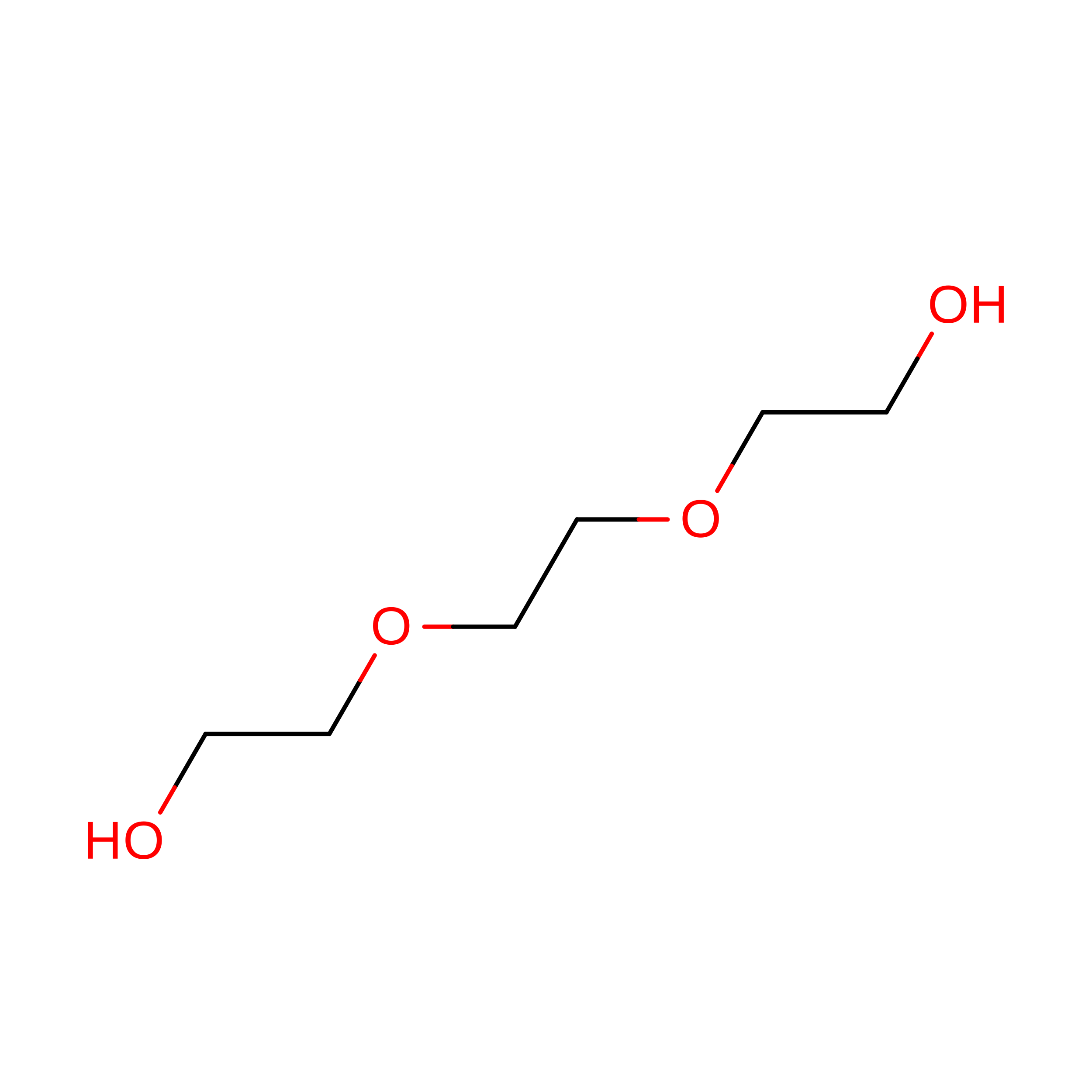
TRIETHYLENE GLYCOL
C6 H14 O4
ZIBGPFATKBEMQZ-UHFFFAOYSA-N
PEG

Query on PEG


Download:Ideal Coordinates CCD File
H [auth A]DI(HYDROXYETHYL)ETHER
C4 H10 O3
MTHSVFCYNBDYFN-UHFFFAOYSA-N
PO4

Query on PO4


Download:Ideal Coordinates CCD File
C [auth A]PHOSPHATE ION
O4 P
NBIIXXVUZAFLBC-UHFFFAOYSA-K
Modified Residues  1 Unique
IDChains TypeFormula2D DiagramParent
NRQ
Query on NRQ
A
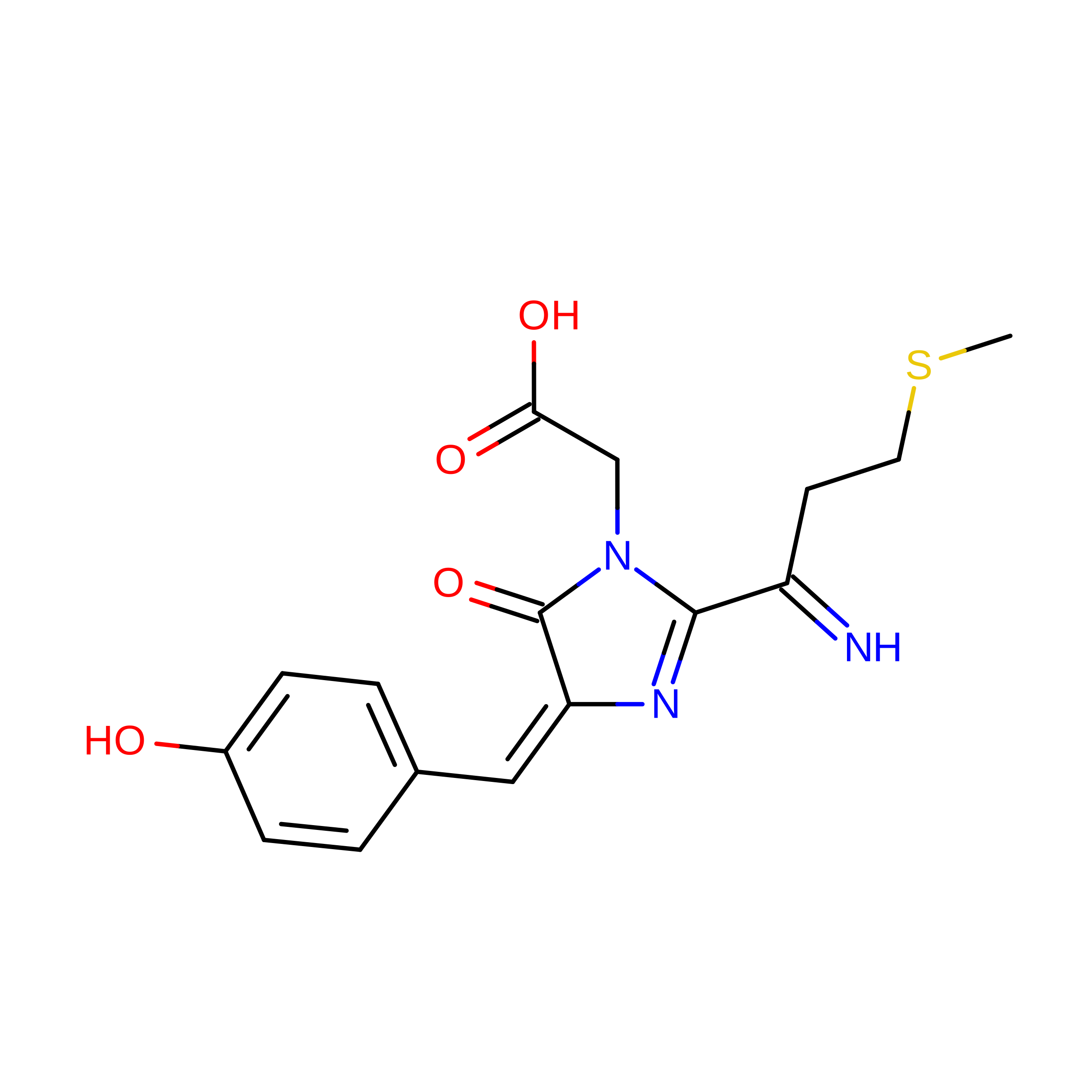
L-PEPTIDE LINKINGC16 H17 N3 O4 SMET, TYR, GLY

Experimental Data & Validation

Experimental Data

  • Method: X-RAY DIFFRACTION
  • Resolution: 1.47 Å
  • R-Value Free:  0.167 (Depositor), 0.167 (DCC) 
  • R-Value Work:  0.137 (Depositor), 0.136 (DCC) 
  • R-Value Observed: 0.138 (Depositor) 
Space Group: C 1 2 1
Unit Cell:
Length ( Å )Angle ( ˚ )
a = 84.2α = 90
b = 35.64β = 109.69
c = 86.01γ = 90
Software Package:
Software NamePurpose
XDSdata reduction
XSCALEdata scaling
PHASERphasing
REFMACrefinement
PDB_EXTRACTdata extraction

Structure Validation

View Full Validation Report


Entry History 

Deposition Data

Revision History  (Full details and data files)

  • Version 1.0: 2016-12-07
    Type: Initial release
  • Version 1.1: 2017-01-11
    Changes: Database references
  • Version 2.0: 2022-09-14
    Changes: Database references, Non-polymer description, Structure summary
  • Version 2.1: 2024-01-31
    Changes: Data collection, Refinement description
  • Version 3.0: 2026-03-18
    Changes: Polymer sequence, Structure summary