Chemistry at the protein-mineral interface in L-ferritin assists the assembly of a functional ( mu (3)-oxo)Tris[( mu (2)-peroxo)] triiron(III) cluster.
Pozzi, C., Ciambellotti, S., Bernacchioni, C., Di Pisa, F., Mangani, S., Turano, P.(2017) Proc Natl Acad Sci U S A 114: 2580-2585
- PubMed: 28202724 Search on PubMedSearch on PubMed Central
- DOI: https://doi.org/10.1073/pnas.1614302114
- Primary Citation Related Structures:
5LG2, 5LG8 - PubMed Abstract:
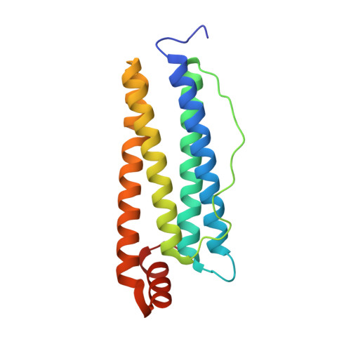
X-ray structures of homopolymeric L-ferritin obtained by freezing protein crystals at increasing exposure times to a ferrous solution showed the progressive formation of a triiron cluster on the inner cage surface of each subunit. After 60 min exposure, a fully assembled (μ 3 -oxo)Tris[(μ 2 -peroxo)(μ 2 -glutamato-κ O :κ O ')](glutamato-κ O )(diaquo)triiron(III) anionic cluster appears in human L-ferritin. Glu60, Glu61, and Glu64 provide the anchoring of the cluster to the protein cage. Glu57 shuttles incoming iron ions toward the cluster. We observed a similar metallocluster in horse spleen L-ferritin, indicating that it represents a common feature of mammalian L-ferritins. The structures suggest a mechanism for iron mineral formation at the protein interface. The functional significance of the observed patch of carboxylate side chains and resulting metallocluster for biomineralization emerges from the lower iron oxidation rate measured in the E60AE61AE64A variant of human L-ferritin, leading to the proposal that the observed metallocluster corresponds to the suggested, but yet unobserved, nucleation site of L-ferritin.
- Department of Biotechnology, Chemistry, and Pharmacy, University of Siena, Siena 53100, Italy.
Organizational Affiliation: