Membrane Interactions of the Mason-Pfizer Monkey Virus Matrix Protein and Its Budding Deficient Mutants.
Kroupa, T., Langerova, H., Dolezal, M., Prchal, J., Spiwok, V., Hunter, E., Rumlova, M., Hrabal, R., Ruml, T.(2016) J Mol Biology 428: 4708-4722
- PubMed: 27725181 Search on PubMed
- DOI: https://doi.org/10.1016/j.jmb.2016.10.010
- Primary Citation Related Structures:
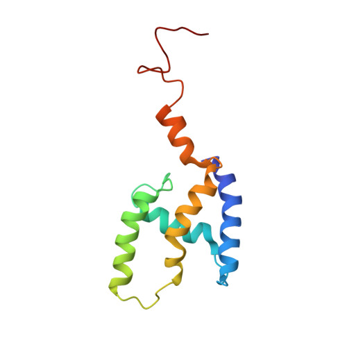
5LDL, 5LMY - PubMed Abstract:
Matrix proteins (MAs) play a key role in the transport of retroviral proteins inside infected cells and in the interaction with cellular membranes. In most retroviruses, retroviral MAs are N-terminally myristoylated. This modification serves as a membrane targeting signal and also as an anchor for membrane interaction. The aim of this work was to characterize the interactions anchoring retroviral MA at the plasma membrane of infected cell. To address this issue, we compared the structures and membrane affinity of the Mason-Pfizer monkey virus (M-PMV) wild-type MA with its two budding deficient double mutants, that is, T41I/T78I and Y28F/Y67F. The structures of the mutants were determined using solution NMR spectroscopy, and their interactions with water-soluble phospholipids were studied. Water-soluble phospholipids are widely used models for studying membrane interactions by solution NMR spectroscopy. However, this approach might lead to artificial results due to unnatural hydrophobic interactions. Therefore, we used a new approach based on the measurement of the loss of the 1 H NMR signal intensity of the protein sample induced by the addition of the liposomes containing phospholipids with naturally long fatty acids. HIV-1 MA was used as a positive control because its ability to interact with liposomes has already been described. We found that in contrast to HIV-1, the M-PMV MA interacted with the liposomes differently and much weaker. In our invivo experiments, the M-PMV MA did not co-localize with lipid rafts. Therefore, we concluded that M-PMV might adopt a different membrane binding mechanism than HIV-1.
- Department of Biochemistry and Microbiology, University of Chemistry and Technology, Prague, Technická 5, 166 28 Prague 6, Czech Republic; Laboratory of NMR Spectroscopy, University of Chemistry and Technology, Prague, Technická 5, 166 28 Prague 6, Czech Republic. Electronic address: kroupat@vscht.cz.
Organizational Affiliation: