5LAX

Crystal structure of HLA_DRB1*04:01 in complex with alpha-enolase peptide 26-40


Experimental Data Snapshot

  • Method: X-RAY DIFFRACTION
  • Resolution: 2.60 Å
  • R-Value Free: 0.270 
  • R-Value Work: 0.191 
  • R-Value Observed: 0.195 

wwPDB Validation   3D Report Full Report


This is version 1.1 of the entry. See complete history


Literature

Functional and Structural Characterization of a Novel HLA-DRB1*04:01-Restricted alpha-Enolase T Cell Epitope in Rheumatoid Arthritis.

Gerstner, C.Dubnovitsky, A.Sandin, C.Kozhukh, G.Uchtenhagen, H.James, E.A.Ronnelid, J.Ytterberg, A.J.Pieper, J.Reed, E.Tandre, C.Rieck, M.Zubarev, R.A.Ronnblom, L.Sandalova, T.Buckner, J.H.Achour, A.Malmstrom, V.

(2016) Front Immunol 7: 494-494

  • DOI: https://doi.org/10.3389/fimmu.2016.00494
  • Primary Citation of Related Structures:  
    5JLZ, 5LAX

  • PubMed Abstract: 

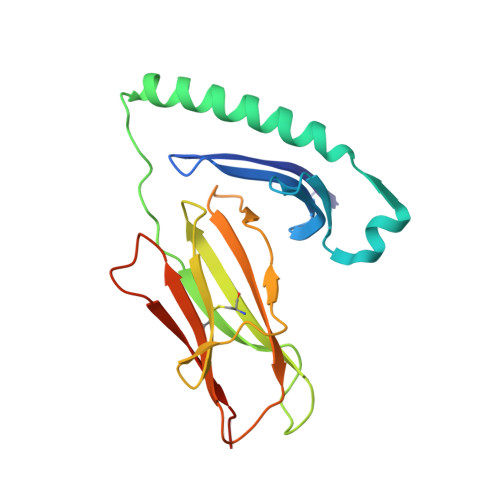
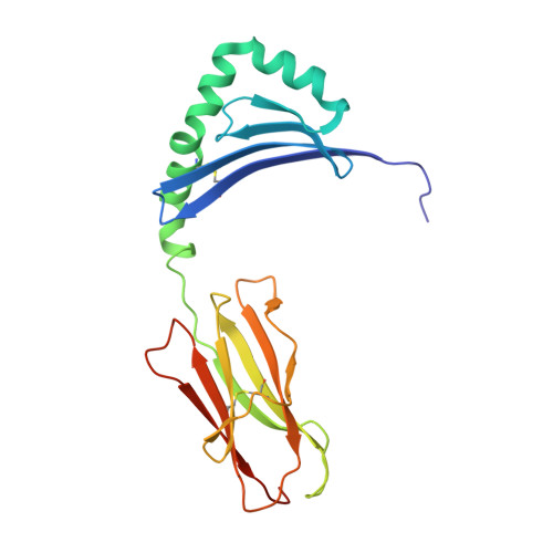
    Antibodies to citrullinated proteins, common in rheumatoid arthritis (RA) patients, are strongly associated to a specific set of HLA-DR alleles including HLA-DRB1*04:01, *04:04, and *01:01. Here, we first demonstrate that autoantibody levels toward the dominant citrullinated B cell epitope from α-enolase are significantly elevated in HLA-DRB1*04:01-positive RA patients. Furthermore, we identified α-enolase-derived T cell epitopes and demonstrated that native and citrullinated versions of several peptides bind with different affinities to HLA-DRB1*04:01, *04:04, and *01:01. The citrulline residues in the eight identified peptides are distributed throughout the entire length of the presented epitopes and more specifically, localized at peptide positions p-2, p2, p4, p6, p7, p10, and p11. Importantly, in contrast to its native version peptide 26 (TSKGLF R AAVPSGAS), the HLA-DRB1*04:01-restricted citrullinated peptide Cit26 (TSKGLF Cit AAVPSGAS) elicited significant functional T cell responses in primary cells from RA patients. Comparative analysis of the crystal structures of HLA-DRB1*04:01 in complex with peptide 26 or Cit26 demonstrated that the posttranslational modification did not alter the conformation of the peptide. And since citrullination is the only structural difference between the two complexes, this indicates that the neo-antigen Cit26 is recognized by T cells with high specificity to the citrulline residue.


  • Organizational Affiliation

    Rheumatology Unit, Department of Medicine Solna, Center for Molecular Medicine, Karolinska Institutet, Karolinska University Hospital , Stockholm , Sweden.


Macromolecules
Find similar proteins by:  (by identity cutoff)  |  3D Structure
Entity ID: 1
MoleculeChains Sequence LengthOrganismDetailsImage
HLA class II histocompatibility antigen, DR alpha chain
A, C
189Homo sapiensMutation(s): 0 
Gene Names: HLA-DRAHLA-DRA1
UniProt & NIH Common Fund Data Resources
Find proteins for P01903 (Homo sapiens)
Explore P01903 
Go to UniProtKB:  P01903
PHAROS:  P01903
GTEx:  ENSG00000204287 
Entity Groups  
Sequence Clusters30% Identity50% Identity70% Identity90% Identity95% Identity100% Identity
UniProt GroupP01903
Sequence Annotations
Expand
  • Reference Sequence
Find similar proteins by:  (by identity cutoff)  |  3D Structure
Entity ID: 2
MoleculeChains Sequence LengthOrganismDetailsImage
HLA class II histocompatibility antigen, DRB1-4 beta chain
B, D
198Homo sapiensMutation(s): 0 
Gene Names: HLA-DRB1
UniProt & NIH Common Fund Data Resources
Find proteins for P01911 (Homo sapiens)
Explore P01911 
Go to UniProtKB:  P01911
PHAROS:  P01911
GTEx:  ENSG00000196126 
Entity Groups  
Sequence Clusters30% Identity50% Identity70% Identity90% Identity95% Identity100% Identity
UniProt GroupP01911
Sequence Annotations
Expand
  • Reference Sequence

Find similar proteins by:  Sequence   |   3D Structure  

Entity ID: 3
MoleculeChains Sequence LengthOrganismDetailsImage
alpha-enolase peptideTSKGLFRAAVPSGAS
E, F
15Homo sapiensMutation(s): 0 
UniProt & NIH Common Fund Data Resources
Find proteins for P06733 (Homo sapiens)
Explore P06733 
Go to UniProtKB:  P06733
PHAROS:  P06733
GTEx:  ENSG00000074800 
Entity Groups  
Sequence Clusters30% Identity50% Identity70% Identity90% Identity95% Identity100% Identity
UniProt GroupP06733
Sequence Annotations
Expand
  • Reference Sequence
Small Molecules
Ligands 1 Unique
IDChains Name / Formula / InChI Key2D Diagram3D Interactions
MLA
Query on MLA

Download Ideal Coordinates CCD File 
G [auth D]MALONIC ACID
C3 H4 O4
OFOBLEOULBTSOW-UHFFFAOYSA-N
Experimental Data & Validation

Experimental Data

  • Method: X-RAY DIFFRACTION
  • Resolution: 2.60 Å
  • R-Value Free: 0.270 
  • R-Value Work: 0.191 
  • R-Value Observed: 0.195 
  • Space Group: P 21 21 21
Unit Cell:
Length ( Å )Angle ( ˚ )
a = 69.176α = 90
b = 73.45β = 90
c = 144.6γ = 90
Software Package:
Software NamePurpose
REFMACrefinement
XDSdata reduction
XDSdata scaling
PHASERphasing

Structure Validation

View Full Validation Report


Entry History 

Deposition Data

Revision History  (Full details and data files)

  • Version 1.0: 2016-12-07
    Type: Initial release
  • Version 1.1: 2024-01-10
    Changes: Data collection, Database references, Refinement description