5LA6

Tubulin-pironetin complex


Experimental Data Snapshot

  • Method: X-RAY DIFFRACTION
  • Resolution: 2.10 Å
  • R-Value Free: 0.231 
  • R-Value Work: 0.184 
  • R-Value Observed: 0.187 

wwPDB Validation   3D Report Full Report


Ligand Structure Quality Assessment 


This is version 1.3 of the entry. See complete history


Literature

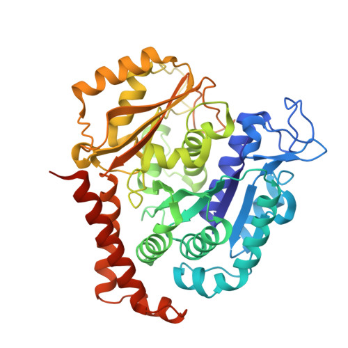
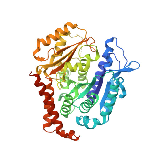
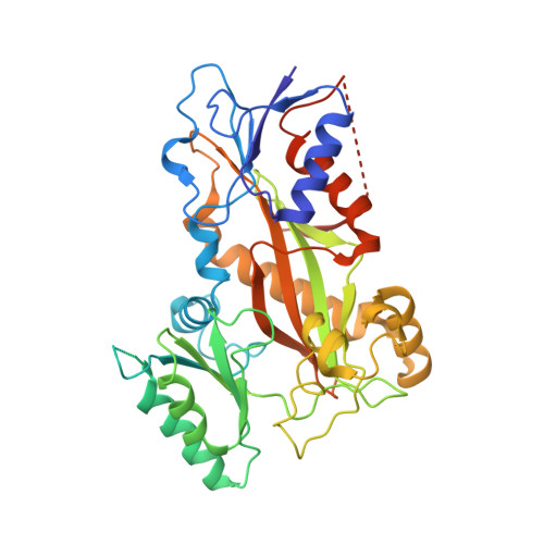
Pironetin Binds Covalently to alpha Cys316 and Perturbs a Major Loop and Helix of alpha-Tubulin to Inhibit Microtubule Formation.

Prota, A.E.Setter, J.Waight, A.B.Bargsten, K.Murga, J.Diaz, J.F.Steinmetz, M.O.

(2016) J Mol Biol 428: 2981-2988

  • DOI: https://doi.org/10.1016/j.jmb.2016.06.023
  • Primary Citation of Related Structures:  
    5LA6

  • PubMed Abstract: 

    Microtubule-targeting agents are among the most powerful drugs used in chemotherapy to treat cancer patients. Pironetin is a natural product that displays promising anticancer properties by binding to and potently inhibiting tubulin assembly into microtubules; however, its molecular mechanism of action remained obscure. Here, we solved the crystal structure of the tubulin-pironetin complex and found that the compound covalently binds to Cys316 of α-tubulin. The structure further revealed that pironetin perturbs the T7 loop and helix H8 of α-tubulin. Since both these elements are essential for establishing longitudinal tubulin contacts in microtubules, this result explains how pironetin inhibits the formation of microtubules. Together, our data define the molecular details of the pironetin binding site on α-tubulin and thus offer a promising basis for the rational design of pironetin variants with improved activity profiles. They further extend our knowledge on strategies evolved by natural products to target and perturb the microtubule cytoskeleton.


  • Organizational Affiliation

    Laboratory of Biomolecular Research, Department of Biology and Chemistry, Paul Scherrer Institut, 5232 Villigen, Switzerland. Electronic address: andrea.prota@psi.ch.


Macromolecules
Find similar proteins by:  (by identity cutoff)  |  3D Structure
Entity ID: 1
MoleculeChains Sequence LengthOrganismDetailsImage
Tubulin alpha-1B chain
A, C
451Bos taurusMutation(s): 0 
UniProt
Find proteins for P81947 (Bos taurus)
Explore P81947 
Go to UniProtKB:  P81947
Entity Groups  
Sequence Clusters30% Identity50% Identity70% Identity90% Identity95% Identity100% Identity
UniProt GroupP81947
Sequence Annotations
Expand
  • Reference Sequence
Find similar proteins by:  (by identity cutoff)  |  3D Structure
Entity ID: 2
MoleculeChains Sequence LengthOrganismDetailsImage
Tubulin beta-2B chain
B, D
445Bos taurusMutation(s): 0 
UniProt
Find proteins for Q6B856 (Bos taurus)
Explore Q6B856 
Go to UniProtKB:  Q6B856
Entity Groups  
Sequence Clusters30% Identity50% Identity70% Identity90% Identity95% Identity100% Identity
UniProt GroupQ6B856
Sequence Annotations
Expand
  • Reference Sequence
Find similar proteins by:  (by identity cutoff)  |  3D Structure
Entity ID: 3
MoleculeChains Sequence LengthOrganismDetailsImage
Stathmin-4143Rattus norvegicusMutation(s): 0 
Gene Names: Stmn4
UniProt
Find proteins for P63043 (Rattus norvegicus)
Explore P63043 
Go to UniProtKB:  P63043
Entity Groups  
Sequence Clusters30% Identity50% Identity70% Identity90% Identity95% Identity100% Identity
UniProt GroupP63043
Sequence Annotations
Expand
  • Reference Sequence
Find similar proteins by:  (by identity cutoff)  |  3D Structure
Entity ID: 4
MoleculeChains Sequence LengthOrganismDetailsImage
Tubulin-Tyrosine Ligase384Gallus gallusMutation(s): 0 
Gene Names: TTL
Entity Groups  
Sequence Clusters30% Identity50% Identity70% Identity90% Identity95% Identity100% Identity
Sequence Annotations
Expand
  • Reference Sequence
Small Molecules
Ligands 6 Unique
IDChains Name / Formula / InChI Key2D Diagram3D Interactions
GTP
Query on GTP

Download Ideal Coordinates CCD File 
G [auth A],
N [auth C]
GUANOSINE-5'-TRIPHOSPHATE
C10 H16 N5 O14 P3
XKMLYUALXHKNFT-UUOKFMHZSA-N
ACP
Query on ACP

Download Ideal Coordinates CCD File 
S [auth F]PHOSPHOMETHYLPHOSPHONIC ACID ADENYLATE ESTER
C11 H18 N5 O12 P3
UFZTZBNSLXELAL-IOSLPCCCSA-N
GDP
Query on GDP

Download Ideal Coordinates CCD File 
K [auth B],
R [auth D]
GUANOSINE-5'-DIPHOSPHATE
C10 H15 N5 O11 P2
QGWNDRXFNXRZMB-UUOKFMHZSA-N
X3H
Query on X3H

Download Ideal Coordinates CCD File 
Q [auth C]PIRONETIN
C19 H34 O4
UKUHYDSKNMYCAN-FSEPSNHWSA-N
CA
Query on CA

Download Ideal Coordinates CCD File 
I [auth A],
J [auth A],
M [auth B],
P [auth C]
CALCIUM ION
Ca
BHPQYMZQTOCNFJ-UHFFFAOYSA-N
MG
Query on MG

Download Ideal Coordinates CCD File 
H [auth A],
L [auth B],
O [auth C]
MAGNESIUM ION
Mg
JLVVSXFLKOJNIY-UHFFFAOYSA-N
Experimental Data & Validation

Experimental Data

  • Method: X-RAY DIFFRACTION
  • Resolution: 2.10 Å
  • R-Value Free: 0.231 
  • R-Value Work: 0.184 
  • R-Value Observed: 0.187 
  • Space Group: P 21 21 21
Unit Cell:
Length ( Å )Angle ( ˚ )
a = 105.68α = 90
b = 157.2β = 90
c = 180.99γ = 90
Software Package:
Software NamePurpose
PHENIXrefinement
XDSdata reduction
XSCALEdata scaling
PHENIXphasing

Structure Validation

View Full Validation Report


Ligand Structure Quality Assessment 


Entry History & Funding Information

Deposition Data


Funding OrganizationLocationGrant Number
Swiss National Science FoundationSwitzerland310030B_138659 and 31003A_166608
Spanish Ministry of Economy and CompetitivenessSpainCTQ2014-52949-P
Cultura i Sport de la Generalitat ValencianaSpainPROMETEO 2013/027
Spanish Ministry of Economy and CompetitivenessSpainBIO2013-42984-R and S2010/BMD-2457 BIPEDD2

Revision History  (Full details and data files)

  • Version 1.0: 2016-07-20
    Type: Initial release
  • Version 1.1: 2016-08-17
    Changes: Database references
  • Version 1.2: 2017-09-13
    Changes: Author supporting evidence
  • Version 1.3: 2024-01-10
    Changes: Data collection, Database references, Derived calculations, Refinement description