5L8H

Structure of USP46-UbVME


Experimental Data Snapshot

  • Method: X-RAY DIFFRACTION
  • Resolution: 1.85 Å
  • R-Value Free: 0.194 
  • R-Value Work: 0.166 
  • R-Value Observed: 0.168 

wwPDB Validation   3D Report Full Report


This is version 2.1 of the entry. See complete history


Literature

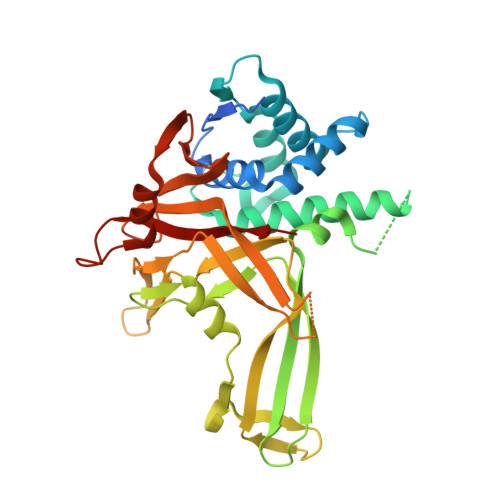
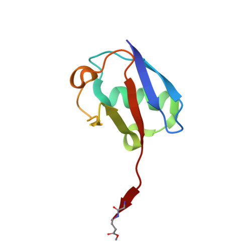
A conserved two-step binding for the UAF1 regulator to the USP12 deubiquitinating enzyme.

Dharadhar, S.Clerici, M.van Dijk, W.J.Fish, A.Sixma, T.K.

(2016) J Struct Biol 196: 437-447

  • DOI: https://doi.org/10.1016/j.jsb.2016.09.011
  • Primary Citation of Related Structures:  
    5L8E, 5L8H, 5L8W

  • PubMed Abstract: 

    Regulation of deubiquitinating enzyme (DUB) activity is an essential step for proper function of cellular ubiquitin signals. UAF1 is a WD40 repeat protein, which binds and activates three important DUBs, USP1, USP12 and USP46. Here, we report the crystal structure of the USP12-Ub/UAF1 complex at a resolution of 2.8Å and of UAF1 at 2.3Å. In the complex we find two potential sites for UAF1 binding, analogous to what was seen in a USP46/UAF1 complex. In line with these observed dual binding states, we show here that USP12/UAF1 complex has 1:2 stoichiometry in solution, with a two-step binding at 4nM and 325nM respectively. Mutagenesis studies show that the fingers sub-domain of USP12 interacts with UAF1 to form the high affinity interface. Our activation studies confirm that the high affinity binding is important for activation while the second UAF1 binding does not affect activation. Nevertheless, we show that this two step binding is conserved in the well-studied USP12 paralog, USP1. Our results highlight the interfaces essential for regulation of USP12 activity and show a conserved second binding of UAF1 which could be important for regulatory functions independent of USP12 activity.


  • Organizational Affiliation

    Division of Biochemistry and CGC.nl, Netherlands Cancer Institute, Plesmanlaan 121, 1066 CX Amsterdam, The Netherlands.


Macromolecules
Find similar proteins by:  (by identity cutoff)  |  3D Structure
Entity ID: 1
MoleculeChains Sequence LengthOrganismDetailsImage
Ubiquitin carboxyl-terminal hydrolase 46359Homo sapiensMutation(s): 0 
Gene Names: USP46
EC: 3.4.19.12
UniProt & NIH Common Fund Data Resources
Find proteins for P62068 (Homo sapiens)
Explore P62068 
Go to UniProtKB:  P62068
PHAROS:  P62068
GTEx:  ENSG00000109189 
Entity Groups  
Sequence Clusters30% Identity50% Identity70% Identity90% Identity95% Identity100% Identity
UniProt GroupP62068
Sequence Annotations
Expand
  • Reference Sequence
Find similar proteins by:  (by identity cutoff)  |  3D Structure
Entity ID: 2
MoleculeChains Sequence LengthOrganismDetailsImage
Polyubiquitin-B76Homo sapiensMutation(s): 1 
Gene Names: UBB
UniProt & NIH Common Fund Data Resources
Find proteins for P0CG47 (Homo sapiens)
Explore P0CG47 
Go to UniProtKB:  P0CG47
PHAROS:  P0CG47
GTEx:  ENSG00000170315 
Entity Groups  
Sequence Clusters30% Identity50% Identity70% Identity90% Identity95% Identity100% Identity
UniProt GroupP0CG47
Sequence Annotations
Expand
  • Reference Sequence
Small Molecules
Ligands 1 Unique
IDChains Name / Formula / InChI Key2D Diagram3D Interactions
ZN
Query on ZN

Download Ideal Coordinates CCD File 
C [auth A]ZINC ION
Zn
PTFCDOFLOPIGGS-UHFFFAOYSA-N
Experimental Data & Validation

Experimental Data

  • Method: X-RAY DIFFRACTION
  • Resolution: 1.85 Å
  • R-Value Free: 0.194 
  • R-Value Work: 0.166 
  • R-Value Observed: 0.168 
  • Space Group: C 2 2 21
Unit Cell:
Length ( Å )Angle ( ˚ )
a = 91.95α = 90
b = 104.66β = 90
c = 135.31γ = 90
Software Package:
Software NamePurpose
REFMACrefinement
XDSdata reduction
XSCALEdata scaling
PHASERphasing

Structure Validation

View Full Validation Report


Entry History & Funding Information

Deposition Data


Funding OrganizationLocationGrant Number
KWFNetherlandsNKI-2014-6858
KWFNetherlandsNKI- 2008-4014
European Research CouncilNetherlands249997

Revision History  (Full details and data files)

  • Version 1.0: 2016-09-28
    Type: Initial release
  • Version 1.1: 2016-11-30
    Changes: Database references
  • Version 2.0: 2023-11-15
    Changes: Atomic model, Author supporting evidence, Data collection, Database references, Derived calculations
  • Version 2.1: 2024-01-10
    Changes: Refinement description