5L6E

Crystal structure of the human METTL3-METTL14 complex bound to SAM


Experimental Data Snapshot

  • Method: X-RAY DIFFRACTION
  • Resolution: 1.90 Å
  • R-Value Free: 0.183 
  • R-Value Work: 0.157 
  • R-Value Observed: 0.159 

wwPDB Validation   3D Report Full Report


Ligand Structure Quality Assessment 


This is version 1.1 of the entry. See complete history


Literature

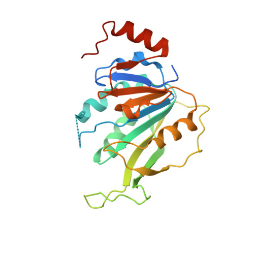
Structural insights into the molecular mechanism of the m(6)A writer complex.

Sledz, P.Jinek, M.

(2016) Elife 5

  • DOI: 10.7554/eLife.18434
  • Primary Citation of Related Structures:  
    5L6D, 5L6E

  • PubMed Abstract: 
  • Methylation of adenosines at the N(6) position (m(6)A) is a dynamic and abundant epitranscriptomic mark that regulates critical aspects of eukaryotic RNA metabolism in numerous biological processes. The RNA methyltransferases METTL3 and METTL14 are components of a multisubunit m(6)A writer complex whose enzymatic activity is substantially higher than the activities of METTL3 or METTL14 alone ...

    Methylation of adenosines at the N(6) position (m(6)A) is a dynamic and abundant epitranscriptomic mark that regulates critical aspects of eukaryotic RNA metabolism in numerous biological processes. The RNA methyltransferases METTL3 and METTL14 are components of a multisubunit m(6)A writer complex whose enzymatic activity is substantially higher than the activities of METTL3 or METTL14 alone. The molecular mechanism underpinning this synergistic effect is poorly understood. Here we report the crystal structure of the catalytic core of the human m(6)A writer complex comprising METTL3 and METTL14. The structure reveals the heterodimeric architecture of the complex and donor substrate binding by METTL3. Structure-guided mutagenesis indicates that METTL3 is the catalytic subunit of the complex, whereas METTL14 has a degenerate active site and plays non-catalytic roles in maintaining complex integrity and substrate RNA binding. These studies illuminate the molecular mechanism and evolutionary history of eukaryotic m(6)A modification in post-transcriptional genome regulation.


    Organizational Affiliation

    Department of Biochemistry, University of Zurich, Zurich, Switzerland.


Macromolecules
Find similar proteins by:  (by identity cutoff)  |  3D Structure
Entity ID: 1
MoleculeChainsSequence LengthOrganismDetailsImage
N6-adenosine-methyltransferase 70 kDa subunitA227Homo sapiensMutation(s): 0 
Gene Names: METTL3MTA70
EC: 2.1.1.62 (PDB Primary Data), 2.1.1.348 (UniProt)
UniProt & NIH Common Fund Data Resources
Find proteins for Q86U44 (Homo sapiens)
Explore Q86U44 
Go to UniProtKB:  Q86U44
PHAROS:  Q86U44
Entity Groups  
Sequence Clusters30% Identity50% Identity70% Identity90% Identity95% Identity100% Identity
UniProt GroupQ86U44
Protein Feature View
Expand
  • Reference Sequence
Find similar proteins by:  (by identity cutoff)  |  3D Structure
Entity ID: 2
MoleculeChainsSequence LengthOrganismDetailsImage
N6-adenosine-methyltransferase subunit METTL14B289Homo sapiensMutation(s): 0 
Gene Names: METTL14KIAA1627
EC: 2.1.1.62
UniProt & NIH Common Fund Data Resources
Find proteins for Q9HCE5 (Homo sapiens)
Explore Q9HCE5 
Go to UniProtKB:  Q9HCE5
PHAROS:  Q9HCE5
Entity Groups  
Sequence Clusters30% Identity50% Identity70% Identity90% Identity95% Identity100% Identity
UniProt GroupQ9HCE5
Protein Feature View
Expand
  • Reference Sequence
Small Molecules
Ligands 3 Unique
IDChainsName / Formula / InChI Key2D Diagram3D Interactions
SAM
Query on SAM

Download Ideal Coordinates CCD File 
C [auth A]S-ADENOSYLMETHIONINE
C15 H22 N6 O5 S
MEFKEPWMEQBLKI-FCKMPRQPSA-N
 Ligand Interaction
ACT
Query on ACT

Download Ideal Coordinates CCD File 
E [auth B]ACETATE ION
C2 H3 O2
QTBSBXVTEAMEQO-UHFFFAOYSA-M
 Ligand Interaction
MG
Query on MG

Download Ideal Coordinates CCD File 
D [auth A]MAGNESIUM ION
Mg
JLVVSXFLKOJNIY-UHFFFAOYSA-N
 Ligand Interaction
Experimental Data & Validation

Experimental Data

  • Method: X-RAY DIFFRACTION
  • Resolution: 1.90 Å
  • R-Value Free: 0.183 
  • R-Value Work: 0.157 
  • R-Value Observed: 0.159 
  • Space Group: P 32 2 1
Unit Cell:
Length ( Å )Angle ( ˚ )
a = 63.949α = 90
b = 63.949β = 90
c = 225.858γ = 120
Software Package:
Software NamePurpose
PHENIXrefinement
XDSdata reduction
XDSdata scaling
PHASERphasing

Structure Validation

View Full Validation Report


Ligand Structure Quality Assessment 


Entry History 

Deposition Data

Revision History  (Full details and data files)

  • Version 1.0: 2016-10-12
    Type: Initial release
  • Version 1.1: 2018-04-25
    Changes: Data collection