5L4R | pdb_00005l4r

X-ray structure of the adduct between thaumatin and cisplatin


Experimental Data Snapshot

  • Method: X-RAY DIFFRACTION
  • Resolution: 1.45 Å
  • R-Value Free: 
    0.154 (Depositor), 0.144 (DCC) 
  • R-Value Work: 
    0.112 (Depositor) 
  • R-Value Observed: 
    0.114 (Depositor) 

Starting Model: experimental
View more details

wwPDB Validation 3D Report Full Report

Validation slider image for 5L4R

This is version 1.2 of the entry. See complete history

Literature

Cisplatin-Protein Interactions: Unexpected Drug Binding to N-Terminal Amine and Lysine Side Chains.

Russo Krauss, I.Ferraro, G.Merlino, A.

(2016) Inorg Chem 55: 7814-7816

  • DOI: https://doi.org/10.1021/acs.inorgchem.6b01234
  • Primary Citation Related Structures: 
    5L4R

  • PubMed Abstract: 

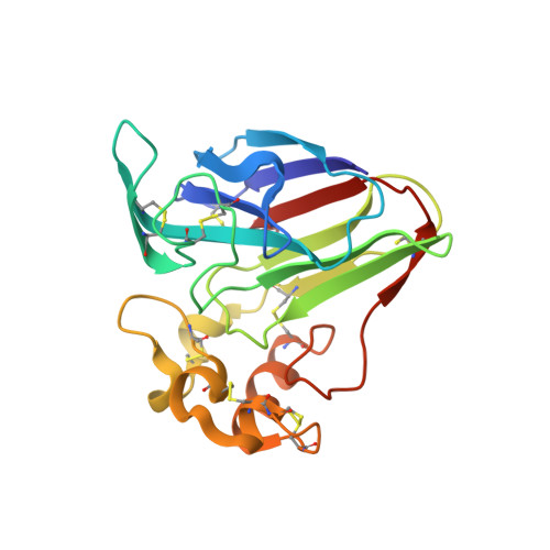
    Literature studies carried out by mass spectrometry and X-ray crystallography have demonstrated that cisplatin is able to bind proteins mainly close to Met, His, and free Cys side chains. To identify possible alternative modes of cisplatin binding to proteins at the molecular level, here we have solved the high-resolution X-ray structure of the adduct formed in the reaction between the drug and the model protein thaumatin, which does not contain any His and free Cys residues and possesses just one buried Met. Our data reveal unexpected cisplatin binding sites on the protein surface that could have general significance: cisplatin fragments -[Pt(NH3)2Cl](+), -[Pt(NH3)Cl2], and -[Pt(NH3)2(OH2)](2+) bind to a protein N-terminus and close to Lys and Glu side chains.


  • Organizational Affiliation
    • Department of Chemical Sciences, University of Naples Federico II , Via Cintia, I-80126 Naples, Italy.

Macromolecule Content 

  • Total Structure Weight: 25.86 kDa 
  • Atom Count: 2,132 
  • Modeled Residue Count: 207 
  • Deposited Residue Count: 207 
  • Unique protein chains: 1

Macromolecules

Find similar proteins by:|  3D Structure
Entity ID: 1
MoleculeChains  Sequence LengthOrganismDetailsImage
Thaumatin-1207Thaumatococcus danielliiMutation(s): 0 
UniProt
Find proteins for P02883 (Thaumatococcus daniellii)
Explore P02883 
Go to UniProtKB:  P02883
Entity Groups
Sequence Clusters30% Identity50% Identity70% Identity90% Identity95% Identity100% Identity
UniProt GroupP02883
Sequence Annotations
Expand
Reference Sequence

Small Molecules

Ligands 4 Unique
IDChains Name / Formula / InChI Key2D Diagram3D Interactions
CPT

Query on CPT


Download:Ideal Coordinates CCD File
E [auth A]
G [auth A]
H [auth A]
I [auth A]
K [auth A]
E [auth A],
G [auth A],
H [auth A],
I [auth A],
K [auth A],
L [auth A],
M [auth A],
N [auth A]
Cisplatin
Cl2 H6 N2 Pt
LXZZYRPGZAFOLE-UHFFFAOYSA-L
73M

Query on 73M


Download:Ideal Coordinates CCD File
D [auth A],
F [auth A],
J [auth A]
bis(azanyl)-chloranyl-oxidanyl-platinum
Cl H5 N2 O Pt
DOKMFIMZADYDDQ-UHFFFAOYSA-L
TLA

Query on TLA


Download:Ideal Coordinates CCD File
B [auth A],
C [auth A]
L(+)-TARTARIC ACID
C4 H6 O6
FEWJPZIEWOKRBE-JCYAYHJZSA-N
GOL

Query on GOL


Download:Ideal Coordinates CCD File
O [auth A]GLYCEROL
C3 H8 O3
PEDCQBHIVMGVHV-UHFFFAOYSA-N

Experimental Data & Validation

Experimental Data

  • Method: X-RAY DIFFRACTION
  • Resolution: 1.45 Å
  • R-Value Free:  0.154 (Depositor), 0.144 (DCC) 
  • R-Value Work:  0.112 (Depositor) 
  • R-Value Observed: 0.114 (Depositor) 
Space Group: P 41 21 2
Unit Cell:
Length ( Å )Angle ( ˚ )
a = 57.837α = 90
b = 57.837β = 90
c = 150.159γ = 90
Software Package:
Software NamePurpose
REFMACrefinement
HKL-2000data reduction
HKL-2000data scaling
PHASERphasing

Structure Validation

View Full Validation Report


Entry History 

Deposition Data

Revision History  (Full details and data files)

  • Version 1.0: 2016-12-07
    Type: Initial release
  • Version 1.1: 2024-01-10
    Changes: Data collection, Database references, Derived calculations, Refinement description
  • Version 1.2: 2024-11-06
    Changes: Structure summary