5L04

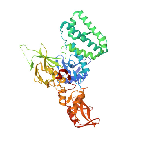
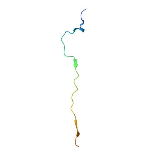
STRUCTURE OF INTERFERON LAMBDA 1 RECEPTOR WITH HUMAN KINASE JAK1


Experimental Data Snapshot

  • Method: X-RAY DIFFRACTION
  • Resolution: 2.10 Å
  • R-Value Free: 0.230 
  • R-Value Work: 0.169 
  • R-Value Observed: 0.171 

wwPDB Validation   3D Report Full Report


This is version 1.2 of the entry. See complete history


Literature

Crystal Structure of a Complex of the Intracellular Domain of Interferon lambda Receptor 1 (IFNLR1) and the FERM/SH2 Domains of Human JAK1.

Zhang, D.Wlodawer, A.Lubkowski, J.

(2016) J Mol Biol 428: 4651-4668

  • DOI: https://doi.org/10.1016/j.jmb.2016.10.005
  • Primary Citation of Related Structures:  
    5L04

  • PubMed Abstract: 

    The crystal structure of a construct consisting of the FERM and SH2-like domains of the human Janus kinase 1 (JAK1) bound to a fragment of the intracellular domain of the interferon-λ receptor 1 (IFNLR1) has been determined at the nominal resolution of 2.1Å. In this structure, the receptor peptide forms an 85-Å-long extended chain, in which both the previously identified box1 and box2 regions bind simultaneously to the FERM and SH2-like domains of JAK1. Both domains of JAK1 are generally well ordered, with regions not seen in the crystal structure limited to loops located away from the receptor-binding regions. The structure provides a much more complete and accurate picture of the interactions between JAK1 and IFNLR1 than those given in earlier reports, illuminating the molecular basis of the JAK-cytokine receptor association. A glutamate residue adjacent to the box2 region in IFNLR1 mimics the mode of binding of a phosphotyrosine in classical SH2 domains. It was shown here that a deletion of residues within the box1 region of the receptor abolishes stable interactions with JAK1, although it was previously shown that box2 alone is sufficient to stabilize a similar complex of the interferon-α receptor and TYK2.


  • Organizational Affiliation

    Macromolecular Crystallography Laboratory, Center for Cancer Research, National Cancer Institute, Frederick, MD 21702, USA.


Macromolecules
Find similar proteins by:  (by identity cutoff)  |  3D Structure
Entity ID: 1
MoleculeChains Sequence LengthOrganismDetailsImage
Tyrosine-protein kinase JAK1550Homo sapiensMutation(s): 0 
Gene Names: JAK1JAK1AJAK1B
EC: 2.7.10.2
UniProt & NIH Common Fund Data Resources
Find proteins for P23458 (Homo sapiens)
Explore P23458 
Go to UniProtKB:  P23458
PHAROS:  P23458
GTEx:  ENSG00000162434 
Entity Groups  
Sequence Clusters30% Identity50% Identity70% Identity90% Identity95% Identity100% Identity
UniProt GroupP23458
Sequence Annotations
Expand
  • Reference Sequence
Find similar proteins by:  (by identity cutoff)  |  3D Structure
Entity ID: 2
MoleculeChains Sequence LengthOrganismDetailsImage
Interferon lambda receptor 148Homo sapiensMutation(s): 0 
Gene Names: IFNLR1IL28RALICR2
UniProt & NIH Common Fund Data Resources
Find proteins for Q8IU57 (Homo sapiens)
Explore Q8IU57 
Go to UniProtKB:  Q8IU57
PHAROS:  Q8IU57
GTEx:  ENSG00000185436 
Entity Groups  
Sequence Clusters30% Identity50% Identity70% Identity90% Identity95% Identity100% Identity
UniProt GroupQ8IU57
Sequence Annotations
Expand
  • Reference Sequence
Experimental Data & Validation

Experimental Data

  • Method: X-RAY DIFFRACTION
  • Resolution: 2.10 Å
  • R-Value Free: 0.230 
  • R-Value Work: 0.169 
  • R-Value Observed: 0.171 
  • Space Group: C 1 2 1
Unit Cell:
Length ( Å )Angle ( ˚ )
a = 165.7α = 90
b = 53.8β = 114.72
c = 87.39γ = 90
Software Package:
Software NamePurpose
SERGUIdata collection
SCALEPACKdata scaling
PHASERphasing
REFMACrefinement
PDB_EXTRACTdata extraction
HKL-3000data reduction

Structure Validation

View Full Validation Report


Entry History 

Deposition Data

Revision History  (Full details and data files)

  • Version 1.0: 2016-10-12
    Type: Initial release
  • Version 1.1: 2017-04-12
    Changes: Database references
  • Version 1.2: 2024-03-06
    Changes: Data collection, Database references