5KYC

Crystal structure of USP7 catalytic domain [V302K] mutant in complex with ubiquitin (malonate bound)


Experimental Data Snapshot

  • Method: X-RAY DIFFRACTION
  • Resolution: 1.43 Å
  • R-Value Free: 0.192 
  • R-Value Work: 0.170 
  • R-Value Observed: 0.171 

wwPDB Validation   3D Report Full Report


This is version 1.2 of the entry. See complete history


Literature

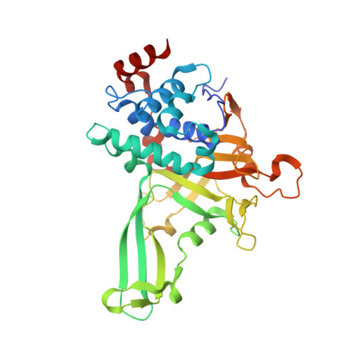
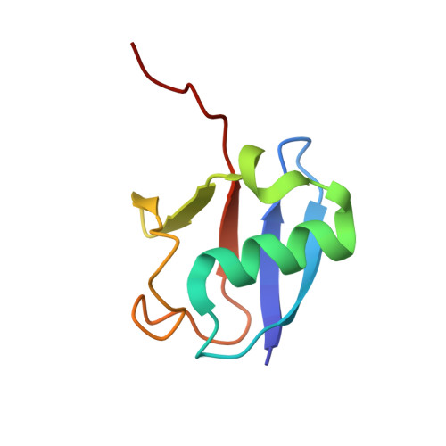
Selectively Modulating Conformational States of USP7 Catalytic Domain for Activation.

Ozen, A.Rouge, L.Bashore, C.Hearn, B.R.Skelton, N.J.Dueber, E.C.

(2018) Structure 26: 72-84.e7

  • DOI: https://doi.org/10.1016/j.str.2017.11.010
  • Primary Citation of Related Structures:  
    5KYB, 5KYC, 5KYD, 5KYE, 5KYF

  • PubMed Abstract: 

    Ubiquitin-specific protease 7 (USP7) deubiquitinase activity is controlled by a number of regulatory factors, including stimulation by intramolecular accessory domains. Alone, the USP7 catalytic domain (USP7cd) shows limited activity and apo USP7cd crystal structures reveal a disrupted catalytic triad. By contrast, ubiquitin-conjugated USP7cd structures demonstrate the canonical cysteine protease active-site geometry; however, the structural features of the USP7cd that stabilize the inactive conformation and the mechanism of transition between inactive and active states remain unclear. Here we use comparative structural analyses, molecular dynamics simulations, and in silico sequence re-engineering via directed sampling by RosettaDesign to identify key molecular determinants of USP7cd activation and successfully engineer USP7cd for improved activity. Full kinetic analysis and multiple X-ray crystal structures of our designs indicate that electrostatic interactions in the distal "switching loop" region and local packing in the hydrophobic core mediate subtle but significant conformational changes that modulate USP7cd activation.


  • Organizational Affiliation

    Department of Early Discovery Biochemistry, Genentech Inc., South San Francisco, CA 94080, USA; Department of Discovery Chemistry, Genentech Inc., South San Francisco, CA 94080, USA.


Macromolecules
Find similar proteins by:  (by identity cutoff)  |  3D Structure
Entity ID: 1
MoleculeChains Sequence LengthOrganismDetailsImage
Ubiquitin carboxyl-terminal hydrolase 7A [auth B]351Homo sapiensMutation(s): 1 
Gene Names: USP7HAUSP
EC: 3.4.19.12
UniProt & NIH Common Fund Data Resources
Find proteins for Q93009 (Homo sapiens)
Explore Q93009 
Go to UniProtKB:  Q93009
PHAROS:  Q93009
GTEx:  ENSG00000187555 
Entity Groups  
Sequence Clusters30% Identity50% Identity70% Identity90% Identity95% Identity100% Identity
UniProt GroupQ93009
Sequence Annotations
Expand
  • Reference Sequence
Find similar proteins by:  (by identity cutoff)  |  3D Structure
Entity ID: 2
MoleculeChains Sequence LengthOrganismDetailsImage
Polyubiquitin-BB [auth C]76Homo sapiensMutation(s): 0 
Gene Names: UBB
UniProt & NIH Common Fund Data Resources
Find proteins for P0CG47 (Homo sapiens)
Explore P0CG47 
Go to UniProtKB:  P0CG47
PHAROS:  P0CG47
GTEx:  ENSG00000170315 
Entity Groups  
Sequence Clusters30% Identity50% Identity70% Identity90% Identity95% Identity100% Identity
UniProt GroupP0CG47
Sequence Annotations
Expand
  • Reference Sequence
Experimental Data & Validation

Experimental Data

  • Method: X-RAY DIFFRACTION
  • Resolution: 1.43 Å
  • R-Value Free: 0.192 
  • R-Value Work: 0.170 
  • R-Value Observed: 0.171 
  • Space Group: C 1 2 1
Unit Cell:
Length ( Å )Angle ( ˚ )
a = 116.33α = 90
b = 43.54β = 107.55
c = 84.76γ = 90
Software Package:
Software NamePurpose
PHENIXrefinement
MOSFLMdata reduction
Aimlessdata scaling
PHASERphasing
PDB_EXTRACTdata extraction

Structure Validation

View Full Validation Report


Entry History 

Deposition Data

Revision History  (Full details and data files)

  • Version 1.0: 2017-08-09
    Type: Initial release
  • Version 1.1: 2019-02-20
    Changes: Data collection, Database references
  • Version 1.2: 2023-10-04
    Changes: Data collection, Database references, Refinement description