5KSU

Crystal structure of HLA-DQ2.5-CLIP1 at 2.73 resolution


Experimental Data Snapshot

  • Method: X-RAY DIFFRACTION
  • Resolution: 2.73 Å
  • R-Value Free: 0.249 
  • R-Value Work: 0.190 
  • R-Value Observed: 0.193 

wwPDB Validation   3D Report Full Report


This is version 1.1 of the entry. See complete history


Literature

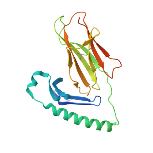
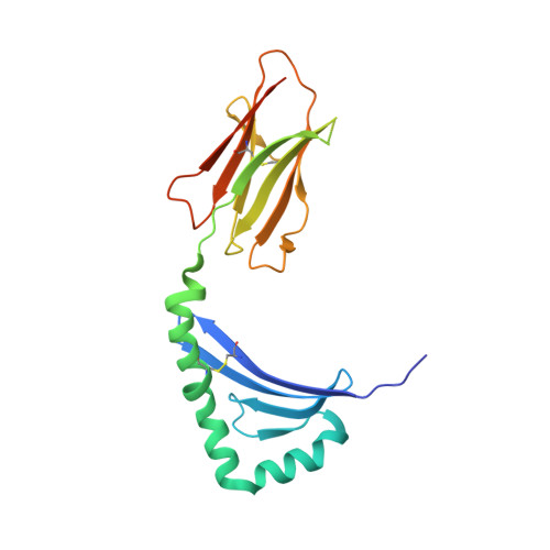
Unraveling the structural basis for the unusually rich association of human leukocyte antigen DQ2.5 with class-II-associated invariant chain peptides.

Nguyen, T.B.Jayaraman, P.Bergseng, E.Madhusudhan, M.S.Kim, C.Y.Sollid, L.M.

(2017) J Biol Chem 292: 9218-9228

  • DOI: https://doi.org/10.1074/jbc.M117.785139
  • Primary Citation of Related Structures:  
    5KSU, 5KSV

  • PubMed Abstract: 
  • Human leukocyte antigen (HLA)-DQ2.5 ( DQA1 * 05/DQB1 * 02 ) is a class-II major histocompatibility complex protein associated with both type 1 diabetes and celiac disease. One unusual feature of DQ2.5 is its high class-II-associated invariant chain peptide (CLIP) content ...

    Human leukocyte antigen (HLA)-DQ2.5 ( DQA1 * 05/DQB1 * 02 ) is a class-II major histocompatibility complex protein associated with both type 1 diabetes and celiac disease. One unusual feature of DQ2.5 is its high class-II-associated invariant chain peptide (CLIP) content. Moreover, HLA-DQ2.5 preferentially binds the non-canonical CLIP2 over the canonical CLIP1. To better understand the structural basis of HLA-DQ2.5's unusual CLIP association characteristics, better insight into the HLA-DQ2.5·CLIP complex structures is required. To this end, we determined the X-ray crystal structure of the HLA-DQ2.5· CLIP1 and HLA-DQ2.5·CLIP2 complexes at 2.73 and 2.20 Å, respectively. We found that HLA-DQ2.5 has an unusually large P4 pocket and a positively charged peptide-binding groove that together promote preferential binding of CLIP2 over CLIP1. An α9-α22-α24-α31-β86-β90 hydrogen bond network located at the bottom of the peptide-binding groove, spanning from the P1 to P4 pockets, renders the residues in this region relatively immobile. This hydrogen bond network, along with a deletion mutation at α53, may lead to HLA-DM insensitivity in HLA-DQ2.5. A molecular dynamics simulation experiment reported here and recent biochemical studies by others support this hypothesis. The diminished HLA-DM sensitivity is the likely reason for the CLIP-rich phenotype of HLA-DQ2.5.


    Organizational Affiliation

    the Centre for Immune Regulation and Department of Immunology, University of Oslo and Oslo University Hospital, N-0372 Oslo, Norway l.m.sollid@medisin.uio.no.


Macromolecules
Find similar proteins by:  (by identity cutoff)  |  3D Structure
Entity ID: 1
MoleculeChainsSequence LengthOrganismDetailsImage
HLA class II histocompatibility antigen, DQ alpha 1 chain
A, D
199Homo sapiensMutation(s): 0 
Gene Names: HLA-DQA1
UniProt & NIH Common Fund Data Resources
Find proteins for P01909 (Homo sapiens)
Explore P01909 
Go to UniProtKB:  P01909
PHAROS:  P01909
Entity Groups  
Sequence Clusters30% Identity50% Identity70% Identity90% Identity95% Identity100% Identity
UniProt GroupP01909
Protein Feature View
Expand
  • Reference Sequence
Find similar proteins by:  (by identity cutoff)  |  3D Structure
Entity ID: 2
MoleculeChainsSequence LengthOrganismDetailsImage
MHC class II HLA-DQ-beta-1
B, E
204Homo sapiensMutation(s): 0 
Gene Names: HLA-DQB1
Entity Groups  
Sequence Clusters30% Identity50% Identity70% Identity90% Identity95% Identity100% Identity
Protein Feature View
Expand
  • Reference Sequence

Find similar proteins by:  Sequence   |   3D Structure  

Entity ID: 3
MoleculeChainsSequence LengthOrganismDetailsImage
HLA class II histocompatibility antigen gamma chain
C, F
15Homo sapiensMutation(s): 0 
Gene Names: CD74DHLAG
UniProt & NIH Common Fund Data Resources
Find proteins for P04233 (Homo sapiens)
Explore P04233 
Go to UniProtKB:  P04233
PHAROS:  P04233
Entity Groups  
Sequence Clusters30% Identity50% Identity70% Identity90% Identity95% Identity100% Identity
UniProt GroupP04233
Protein Feature View
Expand
  • Reference Sequence
Experimental Data & Validation

Experimental Data

  • Method: X-RAY DIFFRACTION
  • Resolution: 2.73 Å
  • R-Value Free: 0.249 
  • R-Value Work: 0.190 
  • R-Value Observed: 0.193 
  • Space Group: C 1 2 1
Unit Cell:
Length ( Å )Angle ( ˚ )
a = 128.86α = 90
b = 69.21β = 110.29
c = 146.69γ = 90
Software Package:
Software NamePurpose
PHENIXrefinement
HKL-2000data reduction
XSCALEdata scaling
PHASERphasing

Structure Validation

View Full Validation Report


Entry History 

Deposition Data

Revision History  (Full details and data files)

  • Version 1.0: 2017-04-05
    Type: Initial release
  • Version 1.1: 2018-04-18
    Changes: Data collection, Database references