5KQ4

Crystal structure of S. pombe Dcp1/Dcp2 in complex with H. sapiens PNRC2 and synthetic cap analog


Experimental Data Snapshot

  • Method: X-RAY DIFFRACTION
  • Resolution: 2.56 Å
  • R-Value Free: 0.251 
  • R-Value Work: 0.206 
  • R-Value Observed: 0.209 

wwPDB Validation   3D Report Full Report


Ligand Structure Quality Assessment 


This is version 1.6 of the entry. See complete history


Literature

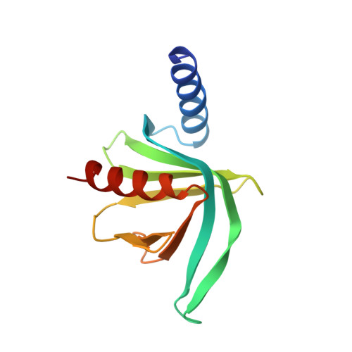
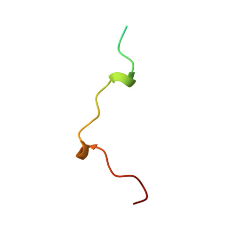
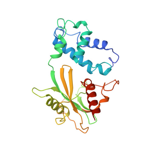
Structural basis of mRNA-cap recognition by Dcp1-Dcp2.

Mugridge, J.S.Ziemniak, M.Jemielity, J.Gross, J.D.

(2016) Nat Struct Mol Biol 23: 987-994

  • DOI: https://doi.org/10.1038/nsmb.3301
  • Primary Citation of Related Structures:  
    5KQ1, 5KQ4

  • PubMed Abstract: 

    Removal of the 5' cap on mRNA by the decapping enzyme Dcp2 is a critical step in 5'-to-3' mRNA decay. Understanding the structural basis of Dcp2 activity has been a challenge because Dcp2 is dynamic and has weak affinity for the cap substrate. Here we present a 2.6-Å-resolution crystal structure of a heterotrimer of fission yeast Dcp2, its essential activator Dcp1, and the human NMD cofactor PNRC2, in complex with a tight-binding cap analog. Cap binding is accompanied by a conformational change in Dcp2, thereby forming a composite nucleotide-binding site comprising conserved residues in the catalytic and regulatory domains. Kinetic analysis of PNRC2 revealed that a conserved short linear motif enhances both substrate affinity and the catalytic step of decapping. These findings explain why Dcp2 requires a conformational change for efficient catalysis and reveals that coactivators promote RNA binding and the catalytic step of decapping, possibly through different conformational states.


  • Organizational Affiliation

    Department of Pharmaceutical Chemistry, University of California, San Francisco, San Francisco, California, USA.


Macromolecules
Find similar proteins by:  (by identity cutoff)  |  3D Structure
Entity ID: 1
MoleculeChains Sequence LengthOrganismDetailsImage
mRNA-decapping enzyme subunit 1A [auth D],
D [auth A]
130Schizosaccharomyces pombe 972h-Mutation(s): 0 
Gene Names: dcp1SPBC3B9.21
UniProt
Find proteins for Q9P805 (Schizosaccharomyces pombe (strain 972 / ATCC 24843))
Explore Q9P805 
Go to UniProtKB:  Q9P805
Entity Groups  
Sequence Clusters30% Identity50% Identity70% Identity90% Identity95% Identity100% Identity
UniProt GroupQ9P805
Sequence Annotations
Expand
  • Reference Sequence
Find similar proteins by:  (by identity cutoff)  |  3D Structure
Entity ID: 2
MoleculeChains Sequence LengthOrganismDetailsImage
Proline-rich nuclear receptor coactivator 2B [auth F],
E [auth C]
31Homo sapiensMutation(s): 0 
Gene Names: PNRC2HSPC208
UniProt & NIH Common Fund Data Resources
Find proteins for Q9NPJ4 (Homo sapiens)
Explore Q9NPJ4 
Go to UniProtKB:  Q9NPJ4
PHAROS:  Q9NPJ4
GTEx:  ENSG00000189266 
Entity Groups  
Sequence Clusters30% Identity50% Identity70% Identity90% Identity95% Identity100% Identity
UniProt GroupQ9NPJ4
Sequence Annotations
Expand
  • Reference Sequence
Find similar proteins by:  (by identity cutoff)  |  3D Structure
Entity ID: 3
MoleculeChains Sequence LengthOrganismDetailsImage
mRNA decapping complex subunit 2C [auth E],
F [auth B]
249Schizosaccharomyces pombe 972h-Mutation(s): 0 
Gene Names: dcp2SPAC19A8.12
EC: 3
UniProt
Find proteins for O13828 (Schizosaccharomyces pombe (strain 972 / ATCC 24843))
Explore O13828 
Go to UniProtKB:  O13828
Entity Groups  
Sequence Clusters30% Identity50% Identity70% Identity90% Identity95% Identity100% Identity
UniProt GroupO13828
Sequence Annotations
Expand
  • Reference Sequence
Small Molecules
Ligands 1 Unique
IDChains Name / Formula / InChI Key2D Diagram3D Interactions
6VQ
Query on 6VQ

Download Ideal Coordinates CCD File 
G [auth E],
H [auth B]
[[(2~{R},3~{S},4~{R},5~{R})-5-(2-azanyl-7-methyl-6-oxidanylidene-3~{H}-purin-7-ium-9-yl)-3,4-bis(oxidanyl)oxolan-2-yl]methoxy-sulfanyl-phosphoryl] [[[(2~{R},3~{S},4~{R},5~{R})-5-(2-azanyl-7-methyl-6-oxidanylidene-3~{H}-purin-7-ium-9-yl)-3,4-bis(oxidanyl)oxolan-2-yl]methoxy-sulfanyl-phosphoryl]oxy-oxidanyl-phosphoryl] hydrogen phosphate
C22 H34 N10 O19 P4 S2
SZQMTBAJCPCVLP-BZDQESSTSA-P
Experimental Data & Validation

Experimental Data

  • Method: X-RAY DIFFRACTION
  • Resolution: 2.56 Å
  • R-Value Free: 0.251 
  • R-Value Work: 0.206 
  • R-Value Observed: 0.209 
  • Space Group: P 1 21 1
Unit Cell:
Length ( Å )Angle ( ˚ )
a = 43.067α = 90
b = 120.752β = 97.9
c = 91.849γ = 90
Software Package:
Software NamePurpose
PHENIXrefinement
Blu-Icedata collection
HKL-2000data reduction
HKL-2000data scaling
PHASERphasing
Cootmodel building
PDB_EXTRACTdata extraction

Structure Validation

View Full Validation Report


Ligand Structure Quality Assessment 


Entry History & Funding Information

Deposition Data


Funding OrganizationLocationGrant Number
National Institutes of Health/National Institute of General Medical Sciences (NIH/NIGMS)United StatesGM078360
National Institutes of Health/National Institute of General Medical Sciences (NIH/NIGMS)United StatesGM105313

Revision History  (Full details and data files)

  • Version 1.0: 2016-10-05
    Type: Initial release
  • Version 1.1: 2016-10-19
    Changes: Database references
  • Version 1.2: 2016-11-16
    Changes: Database references
  • Version 1.3: 2017-09-20
    Changes: Author supporting evidence, Derived calculations
  • Version 1.4: 2017-11-01
    Changes: Author supporting evidence
  • Version 1.5: 2019-12-25
    Changes: Author supporting evidence
  • Version 1.6: 2023-10-04
    Changes: Data collection, Database references, Refinement description