


Crystal structure of lysozyme
Ko, S., Choe, J.To be published.
Experimental Data Snapshot
Starting Model: experimental
View more details
wwPDB Validation 3D Report Full Report

Entity ID: 1 | |||||
|---|---|---|---|---|---|
| Molecule | Chains | Sequence Length | Organism | Details | Image |
| Lysozyme C | 129 | Gallus gallus | Mutation(s): 0 Gene Names: LYZ EC: 3.2.1.17 | 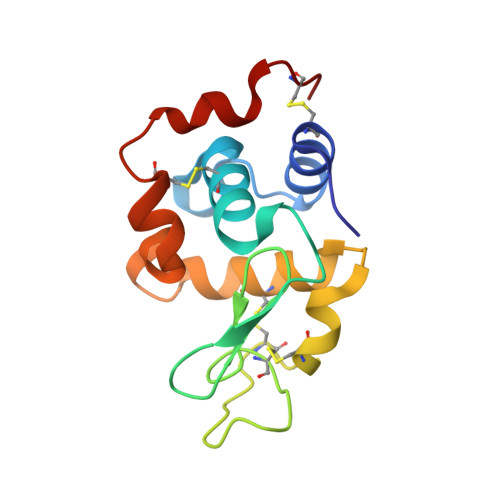 | |
UniProt | |||||
Entity Groups | |||||
| Sequence Clusters | 30% Identity50% Identity70% Identity90% Identity95% Identity100% Identity | ||||
| UniProt Group | P00698 | ||||
Sequence AnnotationsExpand | |||||
Reference Sequence | |||||
| Length ( Å ) | Angle ( ˚ ) |
|---|---|
| a = 76.956 | α = 90 |
| b = 76.956 | β = 90 |
| c = 37.382 | γ = 90 |
| Software Name | Purpose |
|---|---|
| REFMAC | refinement |
| HKL-2000 | data scaling |
| MOLREP | phasing |
| PDB_EXTRACT | data extraction |
| HKL-2000 | data reduction |


