5JWY

Structure of lipid phosphate phosphatase PgpB complex with PE


Experimental Data Snapshot

  • Method: X-RAY DIFFRACTION
  • Resolution: 3.20 Å
  • R-Value Free: 0.305 
  • R-Value Work: 0.267 
  • R-Value Observed: 0.269 

wwPDB Validation   3D Report Full Report


Ligand Structure Quality Assessment 


This is version 1.4 of the entry. See complete history


Literature

Structural Insight into Substrate Selection and Catalysis of Lipid Phosphate Phosphatase PgpB in the Cell Membrane.

Tong, S.Lin, Y.Lu, S.Wang, M.Bogdanov, M.Zheng, L.

(2016) J Biol Chem 291: 18342-18352

  • DOI: https://doi.org/10.1074/jbc.M116.737874
  • Primary Citation of Related Structures:  
    5JWY

  • PubMed Abstract: 

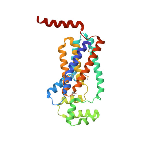
    PgpB belongs to the lipid phosphate phosphatase protein family and is one of three bacterial integral membrane phosphatases catalyzing dephosphorylation of phosphatidylglycerol phosphate (PGP) to generate phosphatidylglycerol. Although the structure of its apo form became recently available, the mechanisms of PgpB substrate binding and catalysis are still unclear. We found that PgpB was inhibited by phosphatidylethanolamine (PE) in a competitive mode in vitro Here we report the crystal structure of the lipid-bound form of PgpB. The structure shows that a PE molecule is stabilized in a membrane-embedded tunnel formed by TM3 and the "PSGH" fingerprint peptide near the catalytic site, providing structural insight into PgpB substrate binding mechanism. Noteworthy, in silico docking of varied lipid phosphates exhibited similar substrate binding modes to that of PE, and the residues in the lipid tunnel appear to be important for PgpB catalysis. The catalytic triad in the active site is essential for dephosphorylating substrates lysophosphatidic acid, phosphatidic acid, or sphingosine-1-phosphate but surprisingly not for the native substrate PGP. Remarkably, residue His-207 alone is sufficient to hydrolyze PGP, indicating a specific catalytic mechanism for PgpB in PG biosynthesis. We also identified two novel sensor residues, Lys-93 and Lys-97, on TM3. Our data show that Lys-97 is essential for the recognition of lyso-form substrates. Modification at the Lys-93 position may alter substrate specificity of lipid phosphate phosphatase proteins in prokaryotes versus eukaryotes. These studies reveal new mechanisms of lipid substrate selection and catalysis by PgpB and suggest that the enzyme rests in a PE-stabilized state in the bilayer.


  • Organizational Affiliation

    From the Center for Membrane Biology, Department of Biochemistry and Molecular Biology, University of Texas Houston Medical School, Houston, Texas 77030 and.


Macromolecules
Find similar proteins by:  (by identity cutoff)  |  3D Structure
Entity ID: 1
MoleculeChains Sequence LengthOrganismDetailsImage
Phosphatidylglycerophosphatase B259Escherichia coli K-12Mutation(s): 0 
Gene Names: pgpBb1278JW1270
EC: 3.1.3.27 (PDB Primary Data), 3.1.3.81 (PDB Primary Data), 3.1.3.4 (PDB Primary Data), 3.6.1.27 (PDB Primary Data)
Membrane Entity: Yes 
UniProt
Find proteins for P0A924 (Escherichia coli (strain K12))
Explore P0A924 
Go to UniProtKB:  P0A924
Entity Groups  
Sequence Clusters30% Identity50% Identity70% Identity90% Identity95% Identity100% Identity
UniProt GroupP0A924
Sequence Annotations
Expand
  • Reference Sequence
Small Molecules
Ligands 1 Unique
IDChains Name / Formula / InChI Key2D Diagram3D Interactions
46E
Query on 46E

Download Ideal Coordinates CCD File 
B [auth A](2R)-3-{[(S)-(2-aminoethoxy)(hydroxy)phosphoryl]oxy}-2-(tetradecanoyloxy)propyl tetradecanoate
C33 H66 N O8 P
NEZDNQCXEZDCBI-WJOKGBTCSA-N
Modified Residues  1 Unique
IDChains TypeFormula2D DiagramParent
MSE
Query on MSE
A
L-PEPTIDE LINKINGC5 H11 N O2 SeMET
Experimental Data & Validation

Experimental Data

  • Method: X-RAY DIFFRACTION
  • Resolution: 3.20 Å
  • R-Value Free: 0.305 
  • R-Value Work: 0.267 
  • R-Value Observed: 0.269 
  • Space Group: P 21 21 21
Unit Cell:
Length ( Å )Angle ( ˚ )
a = 39.533α = 90
b = 73.593β = 90
c = 132.095γ = 90
Software Package:
Software NamePurpose
SCALAdata scaling
REFMACrefinement
PDB_EXTRACTdata extraction
PHENIXphasing

Structure Validation

View Full Validation Report


Ligand Structure Quality Assessment 


Entry History & Funding Information

Deposition Data


Funding OrganizationLocationGrant Number
National Institutes of Health/National Institute of General Medical Sciences (NIH/NIGMS)United StatesGM097290

Revision History  (Full details and data files)

  • Version 1.0: 2016-07-20
    Type: Initial release
  • Version 1.1: 2016-07-27
    Changes: Database references
  • Version 1.2: 2016-09-07
    Changes: Database references
  • Version 1.3: 2017-11-22
    Changes: Database references, Derived calculations, Refinement description
  • Version 1.4: 2019-11-27
    Changes: Author supporting evidence