5JWW

T4 Lysozyme L99A/M102Q with 1-Hydro-2-ethyl-1,2-azaborine Bound


Experimental Data Snapshot

  • Method: X-RAY DIFFRACTION
  • Resolution: 1.47 Å
  • R-Value Free: 0.249 
  • R-Value Work: 0.214 
  • R-Value Observed: 0.216 

wwPDB Validation   3D Report Full Report


This is version 1.3 of the entry. See complete history


Literature

Hydrogen Bonding of 1,2-Azaborines in the Binding Cavity of T4 Lysozyme Mutants: Structures and Thermodynamics.

Lee, H.Fischer, M.Shoichet, B.K.Liu, S.Y.

(2016) J Am Chem Soc 138: 12021-12024

  • DOI: https://doi.org/10.1021/jacs.6b06566
  • Primary Citation of Related Structures:  
    5JWS, 5JWT, 5JWU, 5JWV, 5JWW

  • PubMed Abstract: 

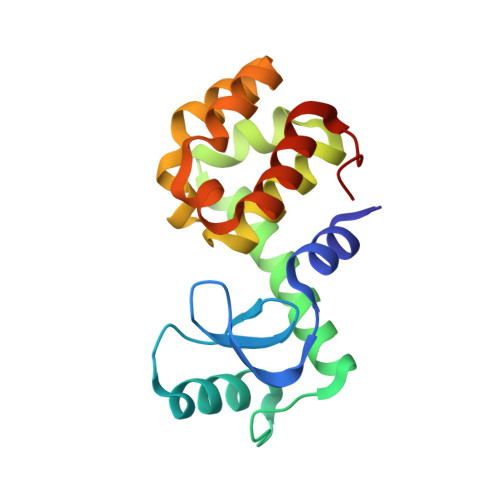
    Protein crystallography and calorimetry were used to characterize the binding of 1,2-azaborines to model cavities in T4 lysozyme in direct comparison to their carbonaceous counterparts. In the apolar L99A cavity, affinity for Ab dropped only slightly versus benzene. In the cavity designed to accommodate a single hydrogen bond (L99A/M102Q), Gln102═O···H-N hydrogen bonding for Ab and BEtAb was observed in the crystallographic complexes. The strength of the hydrogen bonding was estimated as 0.94 and 0.64 kcal/mol for Ab and BEtAb, respectively. This work unambiguously demonstrates that 1,2-azaborines can be readily accommodated in classic aryl recognition pockets and establishes one of 1,2-azaborine's distinguishing features from its carbonaceous isostere benzene: its ability to serve as an NH hydrogen bond donor in a biological setting.


  • Organizational Affiliation

    Department of Chemistry, Boston College , Chestnut Hill, Massachusetts 02467, United States.


Macromolecules
Find similar proteins by:  (by identity cutoff)  |  3D Structure
Entity ID: 1
MoleculeChains Sequence LengthOrganismDetailsImage
Endolysin164Escherichia phage T4Mutation(s): 6 
EC: 3.2.1.17
UniProt
Find proteins for P00720 (Enterobacteria phage T4)
Explore P00720 
Go to UniProtKB:  P00720
Entity Groups  
Sequence Clusters30% Identity50% Identity70% Identity90% Identity95% Identity100% Identity
UniProt GroupP00720
Sequence Annotations
Expand
  • Reference Sequence
Small Molecules
Ligands 2 Unique
IDChains Name / Formula / InChI Key2D Diagram3D Interactions
6OQ
Query on 6OQ

Download Ideal Coordinates CCD File 
C [auth A]2-ethyl-1,2-dihydro-1,2-azaborinine
C6 H10 B N
LWNVLMFFYVOFEQ-UHFFFAOYSA-N
CL
Query on CL

Download Ideal Coordinates CCD File 
B [auth A]CHLORIDE ION
Cl
VEXZGXHMUGYJMC-UHFFFAOYSA-M
Binding Affinity Annotations 
IDSourceBinding Affinity
6OQ Binding MOAD:  5JWW Ka: 2.08e+4 (M^-1) from 1 assay(s)
Experimental Data & Validation

Experimental Data

  • Method: X-RAY DIFFRACTION
  • Resolution: 1.47 Å
  • R-Value Free: 0.249 
  • R-Value Work: 0.214 
  • R-Value Observed: 0.216 
  • Space Group: P 32 2 1
Unit Cell:
Length ( Å )Angle ( ˚ )
a = 60.163α = 90
b = 60.163β = 90
c = 97.087γ = 120
Software Package:
Software NamePurpose
PHENIXrefinement
PDB_EXTRACTdata extraction
d*TREKdata reduction
PHENIXmodel building
PHENIXphasing
d*TREKdata scaling

Structure Validation

View Full Validation Report


Entry History & Funding Information

Deposition Data


Funding OrganizationLocationGrant Number
National Institutes of Health/National Institute of General Medical Sciences (NIH/NIGMS)United States5001989

Revision History  (Full details and data files)

  • Version 1.0: 2016-09-21
    Type: Initial release
  • Version 1.1: 2016-09-28
    Changes: Database references
  • Version 1.2: 2022-04-13
    Changes: Author supporting evidence, Database references, Derived calculations
  • Version 1.3: 2023-09-27
    Changes: Data collection, Refinement description