5JVA | pdb_00005jva

1.95 angstrom crystal structure of TAGRFP-T

  • Classification: FLUORESCENT PROTEIN
  • Organism(s): synthetic construct
  • Mutation(s): No 

  • Deposited: 2016-05-11 Released: 2017-05-17 
  • Deposition Author(s): Hu, X.J.
  • Funding Organization(s): the National Natural Science Foundation of China,

Experimental Data Snapshot

  • Method: X-RAY DIFFRACTION
  • Resolution: 1.95 Å
  • R-Value Free: 
    0.230 (Depositor), 0.237 (DCC) 
  • R-Value Work: 
    0.181 (Depositor), 0.193 (DCC) 
  • R-Value Observed: 
    0.184 (Depositor) 

Starting Model: experimental
View more details

wwPDB Validation 3D Report Full Report

Validation slider image for 5JVA

This is version 2.0 of the entry. See complete history

Literature

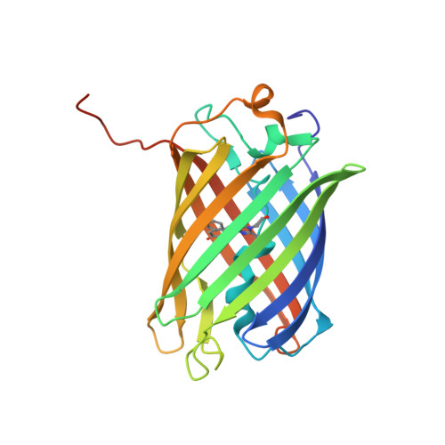
The crystal structure of red fluorescent protein TagRFP-T reveals the mechanism of its superior photostability.

Liu, R.Liang, Q.N.Du, S.Q.Hu, X.J.Ding, Y.

(2016) Biochem Biophys Res Commun 477: 229-234

  • DOI: https://doi.org/10.1016/j.bbrc.2016.06.047
  • Primary Citation Related Structures: 
    5JVA

  • PubMed Abstract: 

    The red fluorescent protein variant TagRFP-T has greatly improved photostability over its parent molecule, TagRFP, but the underlying mechanism leading to this improvement is to date unknown. The 1.95 Å resolution crystallographic structure of TagRFP-T showed that its chromophore exists as a mixture of cis and trans coplanar isomers in roughly equal proportions. Interestingly, both isomers are able to fluoresce, a property that has never been observed in any other fluorescent protein. We propose a "circular restoration model" for TagRFP-T to explain its superior photostability: There are four co-existing chromophore states (cis/trans protonated/ionized state) that can be driven by light to transform from one state into another. This model also explains how TagRPF-T essentially eliminates the temporary dark state (reversible photobleaching).


  • Organizational Affiliation
    • Department of Physiology and Biophysics, School of Life Sciences, Fudan University, Shanghai 200438, China.

Macromolecule Content 

  • Total Structure Weight: 109.12 kDa 
  • Atom Count: 7,730 
  • Modeled Residue Count: 900 
  • Deposited Residue Count: 952 
  • Unique protein chains: 1

Macromolecules

Find similar proteins by:|  3D Structure
Entity ID: 1
MoleculeChains  Sequence LengthOrganismDetailsImage
TagRFP-T
A, B, C, D
238synthetic constructMutation(s): 0 

Experimental Data & Validation

Experimental Data

  • Method: X-RAY DIFFRACTION
  • Resolution: 1.95 Å
  • R-Value Free:  0.230 (Depositor), 0.237 (DCC) 
  • R-Value Work:  0.181 (Depositor), 0.193 (DCC) 
  • R-Value Observed: 0.184 (Depositor) 
Space Group: I 41
Unit Cell:
Length ( Å )Angle ( ˚ )
a = 162.21α = 90
b = 162.21β = 90
c = 76.15γ = 90
Software Package:
Software NamePurpose
REFMACrefinement
HKL-2000data reduction
SCALEPACKdata scaling
MOLREPphasing

Structure Validation

View Full Validation Report


Entry History 

& Funding Information

Deposition Data

  • Released Date: 2017-05-17 
  • Deposition Author(s): Hu, X.J.
  • This entry supersedes: 3T6H

Funding OrganizationLocationGrant Number
the National Natural Science Foundation of China,China31470764

Revision History  (Full details and data files)

  • Version 1.0: 2017-05-17
    Type: Initial release
  • Version 1.1: 2023-11-08
    Changes: Data collection, Database references, Refinement description
  • Version 1.2: 2023-11-15
    Changes: Data collection
  • Version 1.3: 2024-04-03
    Changes: Derived calculations
  • Version 2.0: 2026-03-18
    Changes: Polymer sequence, Structure summary