5JI3

HslUV complex


Experimental Data Snapshot

  • Method: X-RAY DIFFRACTION
  • Resolution: 3.00 Å
  • R-Value Free: 0.253 
  • R-Value Work: 0.222 
  • R-Value Observed: 0.229 

wwPDB Validation   3D Report Full Report


Ligand Structure Quality Assessment 


This is version 1.3 of the entry. See complete history

Re-refinement Note

This entry reflects an alternative modeling of the original data in: 1G4A


Literature

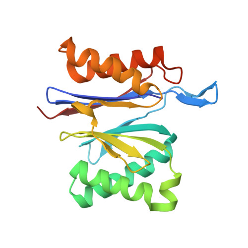
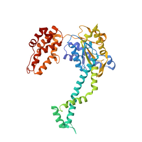
A Structurally Dynamic Region of the HslU Intermediate Domain Controls Protein Degradation and ATP Hydrolysis.

Baytshtok, V.Fei, X.Grant, R.A.Baker, T.A.Sauer, R.T.

(2016) Structure 24: 1766-1777

  • DOI: https://doi.org/10.1016/j.str.2016.08.012
  • Primary Citation of Related Structures:  
    5JI2, 5JI3

  • PubMed Abstract: 

    The I domain of HslU sits above the AAA+ ring and forms a funnel-like entry to the axial pore, where protein substrates are engaged, unfolded, and translocated into HslV for degradation. The L199Q I-domain substitution, which was originally reported as a loss-of-function mutation, resides in a segment that appears to adopt multiple conformations as electron density is not observed in HslU and HslUV crystal structures. The L199Q sequence change does not alter the structure of the AAA+ ring or its interactions with HslV but increases I-domain susceptibility to limited endoproteolysis. Notably, the L199Q mutation increases the rate of ATP hydrolysis substantially, results in slower degradation of some proteins but faster degradation of other substrates, and markedly changes the preference of HslUV for initiating degradation at the N or C terminus of model substrates. Thus, a structurally dynamic region of the I domain plays a key role in controlling protein degradation by HslUV.


  • Organizational Affiliation

    Department of Biology, Massachusetts Institute of Technology, Cambridge, MA 02139, USA.


Macromolecules
Find similar proteins by:  (by identity cutoff)  |  3D Structure
Entity ID: 1
MoleculeChains Sequence LengthOrganismDetailsImage
ATP-dependent protease subunit HslV
A, B, C, D
176Escherichia coliMutation(s): 0 
Gene Names: hslVEC55989_4410
EC: 3.4.25.2
UniProt
Find proteins for P0A7B8 (Escherichia coli (strain K12))
Explore P0A7B8 
Go to UniProtKB:  P0A7B8
Entity Groups  
Sequence Clusters30% Identity50% Identity70% Identity90% Identity95% Identity100% Identity
UniProt GroupP0A7B8
Sequence Annotations
Expand
  • Reference Sequence
Find similar proteins by:  (by identity cutoff)  |  3D Structure
Entity ID: 2
MoleculeChains Sequence LengthOrganismDetailsImage
ATP-dependent protease ATPase subunit HslU
E, F
443Escherichia coliMutation(s): 0 
Gene Names: hslUhtpIZ5478ECs4858
UniProt
Find proteins for P0A6H5 (Escherichia coli (strain K12))
Explore P0A6H5 
Go to UniProtKB:  P0A6H5
Entity Groups  
Sequence Clusters30% Identity50% Identity70% Identity90% Identity95% Identity100% Identity
UniProt GroupP0A6H5
Sequence Annotations
Expand
  • Reference Sequence
Small Molecules
Ligands 1 Unique
IDChains Name / Formula / InChI Key2D Diagram3D Interactions
DAT
Query on DAT

Download Ideal Coordinates CCD File 
G [auth E],
H [auth F]
2'-DEOXYADENOSINE-5'-DIPHOSPHATE
C10 H15 N5 O9 P2
DAEAPNUQQAICNR-RRKCRQDMSA-N
Experimental Data & Validation

Experimental Data

  • Method: X-RAY DIFFRACTION
  • Resolution: 3.00 Å
  • R-Value Free: 0.253 
  • R-Value Work: 0.222 
  • R-Value Observed: 0.229 
  • Space Group: P 3 2 1
Unit Cell:
Length ( Å )Angle ( ˚ )
a = 169.995α = 90
b = 169.995β = 90
c = 161.317γ = 120
Software Package:
Software NamePurpose
CNSrefinement
HKL-2000data reduction
HKL-2000data scaling
CNSphasing
PHENIXrefinement

Structure Validation

View Full Validation Report


Ligand Structure Quality Assessment 


Entry History & Funding Information

Deposition Data


Funding OrganizationLocationGrant Number
National Institutes of HealthUnited States5R01AI016892-36
National Institutes of HealthUnited States5R01GM1011988-37

Revision History  (Full details and data files)

  • Version 1.0: 2016-12-07
    Type: Initial release
  • Version 1.1: 2018-01-31
    Changes: Advisory, Author supporting evidence
  • Version 1.2: 2019-10-16
    Changes: Data collection
  • Version 1.3: 2024-01-10
    Changes: Advisory, Data collection, Database references, Refinement description