5JDT

Structure of Spin-labelled T4 lysozyme mutant L118C-R1 at 100K


Experimental Data Snapshot

  • Method: X-RAY DIFFRACTION
  • Resolution: 1.00 Å
  • R-Value Free: 0.130 
  • R-Value Work: 0.115 
  • R-Value Observed: 0.115 

wwPDB Validation   3D Report Full Report


Ligand Structure Quality Assessment 


This is version 1.2 of the entry. See complete history


Literature

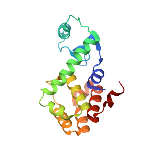
Tracking Transient Conformational States of T4 Lysozyme at Room Temperature Combining X-ray Crystallography and Site-Directed Spin Labeling.

Consentius, P.Gohlke, U.Loll, B.Alings, C.Muller, R.Heinemann, U.Kaupp, M.Wahl, M.Risse, T.

(2016) J Am Chem Soc 138: 12868-12875

  • DOI: https://doi.org/10.1021/jacs.6b05507
  • Primary Citation of Related Structures:  
    5G27, 5JDT

  • PubMed Abstract: 

    Proteins are dynamic molecules that can transiently adopt different conformational states. As the function of the system often depends critically on its conformational state a rigorous understanding of the correlation between structure, energetics and dynamics of the different accessible states is crucial. The biophysical characterization of such processes is, however, challenging as the excited states are often only marginally populated. We show that a combination of X-ray crystallography performed at 100 K as well as at room temperature and EPR spectroscopy on a spin-labeled single crystal allows to correlate the structures of the ground state and a thermally excited state with their thermodynamics using the variant 118R1 of T4 lysozyme as an example. In addition, it is shown that the surrounding solvent can significantly alter the energetic as well as the entropic contribution to the Gibbs free energy without major impact on the structure of both states.


  • Organizational Affiliation

    Institute of Chemistry and Biochemistry, Freie Universität Berlin , Takustr. 3, 14195 Berlin, Germany.


Macromolecules
Find similar proteins by:  (by identity cutoff)  |  3D Structure
Entity ID: 1
MoleculeChains Sequence LengthOrganismDetailsImage
Endolysin164Tequatrovirus T4Mutation(s): 3 
EC: 3.2.1.17
UniProt
Find proteins for P00720 (Enterobacteria phage T4)
Explore P00720 
Go to UniProtKB:  P00720
Entity Groups  
Sequence Clusters30% Identity50% Identity70% Identity90% Identity95% Identity100% Identity
UniProt GroupP00720
Sequence Annotations
Expand
  • Reference Sequence
Small Molecules
Ligands 5 Unique
IDChains Name / Formula / InChI Key2D Diagram3D Interactions
MTN
Query on MTN

Download Ideal Coordinates CCD File 
B [auth A]S-[(1-oxyl-2,2,5,5-tetramethyl-2,5-dihydro-1H-pyrrol-3-yl)methyl] methanesulfonothioate
C10 H18 N O3 S2
MXZPGYFBZHBAQM-UHFFFAOYSA-N
BME
Query on BME

Download Ideal Coordinates CCD File 
F [auth A]BETA-MERCAPTOETHANOL
C2 H6 O S
DGVVWUTYPXICAM-UHFFFAOYSA-N
AZI
Query on AZI

Download Ideal Coordinates CCD File 
G [auth A],
H [auth A]
AZIDE ION
N3
IVRMZWNICZWHMI-UHFFFAOYSA-N
K
Query on K

Download Ideal Coordinates CCD File 
I [auth A]POTASSIUM ION
K
NPYPAHLBTDXSSS-UHFFFAOYSA-N
CL
Query on CL

Download Ideal Coordinates CCD File 
C [auth A],
D [auth A],
E [auth A]
CHLORIDE ION
Cl
VEXZGXHMUGYJMC-UHFFFAOYSA-M
Experimental Data & Validation

Experimental Data

  • Method: X-RAY DIFFRACTION
  • Resolution: 1.00 Å
  • R-Value Free: 0.130 
  • R-Value Work: 0.115 
  • R-Value Observed: 0.115 
  • Space Group: P 32 2 1
Unit Cell:
Length ( Å )Angle ( ˚ )
a = 60.28α = 90
b = 60.28β = 90
c = 96.97γ = 120
Software Package:
Software NamePurpose
PHENIXrefinement
XDSdata reduction
XSCALEdata scaling
PHASERphasing

Structure Validation

View Full Validation Report


Ligand Structure Quality Assessment 


Entry History 

Deposition Data

Revision History  (Full details and data files)

  • Version 1.0: 2016-09-28
    Type: Initial release
  • Version 1.1: 2016-10-26
    Changes: Database references
  • Version 1.2: 2024-01-10
    Changes: Data collection, Database references, Derived calculations, Refinement description