5JBB

Crystal structure of factor IXa variant V16I K98T Y177T I213V in complex with EGR-chloromethylketone


Experimental Data Snapshot

  • Method: X-RAY DIFFRACTION
  • Resolution: 1.56 Å
  • R-Value Free: 0.190 
  • R-Value Work: 0.158 
  • R-Value Observed: 0.160 

wwPDB Validation   3D Report Full Report


Ligand Structure Quality Assessment 


This is version 1.3 of the entry. See complete history


Literature

Releasing the brakes in coagulation Factor IXa by co-operative maturation of the substrate-binding site.

Kristensen, L.H.Olsen, O.H.Blouse, G.E.Brandstetter, H.

(2016) Biochem J 473: 2395-2411

  • DOI: https://doi.org/10.1042/BCJ20160336
  • Primary Citation of Related Structures:  
    5JB8, 5JB9, 5JBA, 5JBB, 5JBC

  • PubMed Abstract: 

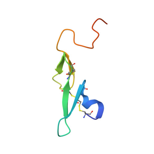
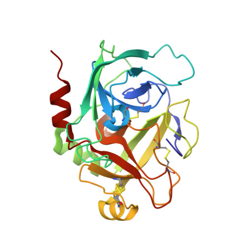
    Coagulation Factor IX is positioned at the merging point of the intrinsic and extrinsic blood coagulation cascades. Factor IXa (activated Factor IX) serves as the trigger for amplification of coagulation through formation of the so-called Xase complex, which is a ternary complex of Factor IXa, its substrate Factor X and the cofactor Factor VIIIa on the surface of activated platelets. Within the Xase complex the substrate turnover by Factor IXa is enhanced 200000-fold; however, the mechanistic and structural basis for this dramatic enhancement remains only partly understood. A multifaceted approach using enzymatic, biophysical and crystallographic methods to evaluate a key set of activity-enhanced Factor IXa variants has demonstrated a delicately balanced bidirectional network. Essential molecular interactions across multiple regions of the Factor IXa molecule co-operate in the maturation of the active site. This maturation is specifically facilitated by long-range communication through the Ile(212)-Ile(213) motif unique to Factor IXa and a flexibility of the 170-loop that is further dependent on the conformation in the Cys(168)-Cys(182) disulfide bond. Ultimately, the network consists of compensatory brakes (Val(16) and Ile(213)) and accelerators (Tyr(99) and Phe(174)) that together allow for a subtle fine-tuning of enzymatic activity.


  • Organizational Affiliation

    Haemophilia Biochemistry, Novo Nordisk A/S, Novo Nordisk Park 1, 2760 Måløv, Denmark Structural Biology Group, Department of Molecular Biology, University of Salzburg, Billrothstrasse 11, 5020 Salzburg, Austria.


Macromolecules
Find similar proteins by:  (by identity cutoff)  |  3D Structure
Entity ID: 1
MoleculeChains Sequence LengthOrganismDetailsImage
Coagulation factor IXA [auth E]58Homo sapiensMutation(s): 0 
Gene Names: F9
EC: 3.4.21.22
UniProt & NIH Common Fund Data Resources
Find proteins for P00740 (Homo sapiens)
Explore P00740 
Go to UniProtKB:  P00740
PHAROS:  P00740
GTEx:  ENSG00000101981 
Entity Groups  
Sequence Clusters30% Identity50% Identity70% Identity90% Identity95% Identity100% Identity
UniProt GroupP00740
Sequence Annotations
Expand
  • Reference Sequence
Find similar proteins by:  (by identity cutoff)  |  3D Structure
Entity ID: 2
MoleculeChains Sequence LengthOrganismDetailsImage
Coagulation factor IXB [auth S]235Homo sapiensMutation(s): 4 
Gene Names: F9
EC: 3.4.21.22
UniProt & NIH Common Fund Data Resources
Find proteins for P00740 (Homo sapiens)
Explore P00740 
Go to UniProtKB:  P00740
PHAROS:  P00740
GTEx:  ENSG00000101981 
Entity Groups  
Sequence Clusters30% Identity50% Identity70% Identity90% Identity95% Identity100% Identity
UniProt GroupP00740
Sequence Annotations
Expand
  • Reference Sequence
Small Molecules
Ligands 3 Unique
IDChains Name / Formula / InChI Key2D Diagram3D Interactions
0GJ
Query on 0GJ

Download Ideal Coordinates CCD File 
D [auth S]L-alpha-glutamyl-N-{(1S)-4-{[amino(iminio)methyl]amino}-1-[(1S)-2-chloro-1-hydroxyethyl]butyl}glycinamide
C14 H28 Cl N6 O5
XELWNHKFCNMWQO-LPEHRKFASA-O
DMS
Query on DMS

Download Ideal Coordinates CCD File 
E [auth S]DIMETHYL SULFOXIDE
C2 H6 O S
IAZDPXIOMUYVGZ-UHFFFAOYSA-N
CA
Query on CA

Download Ideal Coordinates CCD File 
C [auth S]CALCIUM ION
Ca
BHPQYMZQTOCNFJ-UHFFFAOYSA-N
Biologically Interesting Molecules (External Reference) 1 Unique
Experimental Data & Validation

Experimental Data

  • Method: X-RAY DIFFRACTION
  • Resolution: 1.56 Å
  • R-Value Free: 0.190 
  • R-Value Work: 0.158 
  • R-Value Observed: 0.160 
  • Space Group: P 21 21 21
Unit Cell:
Length ( Å )Angle ( ˚ )
a = 44.31α = 90
b = 66.61β = 90
c = 97.05γ = 90
Software Package:
Software NamePurpose
iMOSFLMdata reduction
Aimlessdata scaling
MOLREPphasing
REFMACrefinement
Cootmodel building
PHENIXrefinement
PDB_EXTRACTdata extraction

Structure Validation

View Full Validation Report


Ligand Structure Quality Assessment 


Entry History 

Deposition Data

Revision History  (Full details and data files)

  • Version 1.0: 2016-06-01
    Type: Initial release
  • Version 1.1: 2016-12-21
    Changes: Non-polymer description
  • Version 1.2: 2018-01-24
    Changes: Source and taxonomy
  • Version 1.3: 2024-01-10
    Changes: Data collection, Database references, Derived calculations, Refinement description