Assembly and activation of dynein-dynactin by the cargo adaptor protein Hook3.
Schroeder, C.M., Vale, R.D.(2016) J Cell Biol 214: 309-318
- PubMed: 27482052 Search on PubMedSearch on PubMed Central
- DOI: https://doi.org/10.1083/jcb.201604002
- Primary Citation Related Structures:
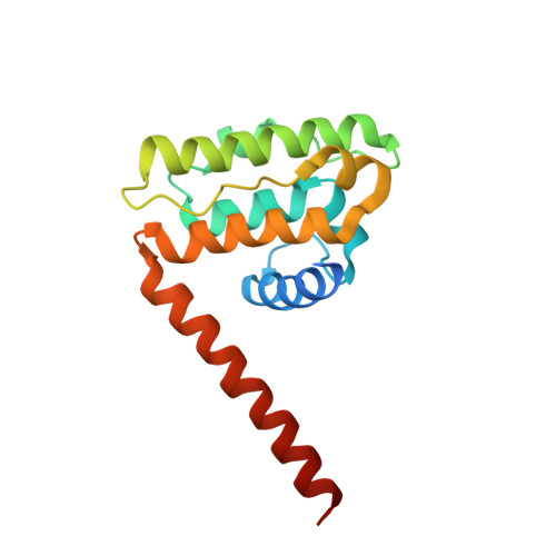
5J8E - PubMed Abstract:
Metazoan cytoplasmic dynein moves processively along microtubules with the aid of dynactin and an adaptor protein that joins dynein and dynactin into a stable ternary complex. Here, we examined how Hook3, a cargo adaptor involved in Golgi and endosome transport, forms a motile dynein-dynactin complex. We show that the conserved Hook domain interacts directly with the dynein light intermediate chain 1 (LIC1). By solving the crystal structure of the Hook domain and using structure-based mutagenesis, we identify two conserved surface residues that are each critical for LIC1 binding. Hook proteins with mutations in these residues fail to form a stable dynein-dynactin complex, revealing a crucial role for LIC1 in this interaction. We also identify a region of Hook3 specifically required for an allosteric activation of processive motility. Our work reveals the structural details of Hook3's interaction with dynein and offers insight into how cargo adaptors form processive dynein-dynactin motor complexes.
- The Howard Hughes Medical Institute, University of California, San Francisco, San Francisco, CA 94158 Department of Cellular and Molecular Pharmacology, University of California, San Francisco, San Francisco, CA 94158.
Organizational Affiliation: